Alternate header for print version


Licensing
Public Domain: This image is in the public domain and thus free of any copyright restrictions. However, as is the norm in scientific publishing and as a matter of courtesy, any user should credit the content provider for any public or private use of this image whenever possible. Learn more
tweet  
*CIL – Cell Image Library accession number. Please use this to reference an image.

CIL:44651*  Cite 
Description

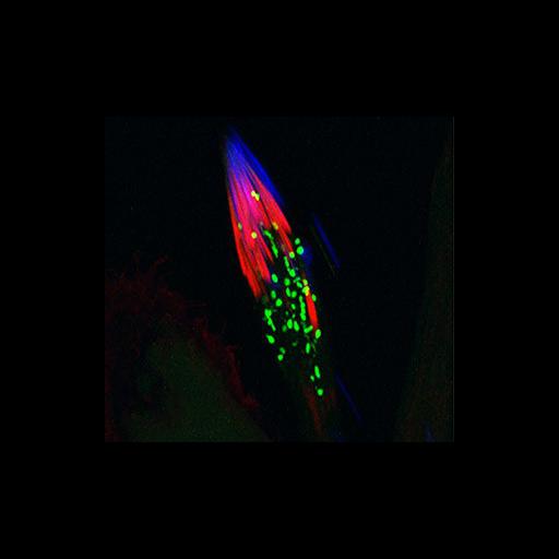
During the differentiation of sperm most cellular proteins and organelles are degraded as they are no longer needed. Actin filaments in the cell help to collect and move cytoplasmic material in maturing sperm, and a cellular chopping machine called the proteasome degrades proteins into amino acids. Shown is a confocal microscopy image of a Drosophila spermatid stained for nuclei (blue), actin filaments (red), and proteasomes (green). A recent study uncovered a role for ADP-ribosylation by tankyrase in the assembly and activity of proteasomes. The image depicting this critical stage of sperm differentiation was selected by the NIGMS for inclusion in the May 2013 issue of Biomedical Beat, a monthly compendium of noteworthy NIGMS-supported research. See also Cho-Park PF and Steller, H 2013. Proteasome regulation by ADP-ribosylation. Cell 153:614-627.

Technical Details

Actin was detected using Alexa 546 phalloidin and proteasomes with transgeneic GFP-alpha6T, a proteosome subunit. Maturing sperm cells in the testis were fixed with formaldehyde, stained, and observed using confocal microscopy with a 40x 1.2 NA objective lens. The image is a projection of a z-stack with step size 1.2 micrometers.

Biological Sources
NCBI Organism Classification
Drosophila melanogaster
Cell Type
spermatid
Cellular Component
cytoskeleton
proteasome complex
Biological Context
Biological Process
spermatogenesis
Attribution
Names
Hermann Steller
Sigi Benjamin-Hong
John M Belote
OTHER
Strang Laboratory of Apoptosis and Cancer Biology, Howard Hughes Medical Institute, The Rockefeller University, 1230 York Avenue, New York, NY 10021, USA.
Citation
Digital Object Identifier (DOI)
doi:10.7295/W9CIL44651
Archival Resource Key (ARK)
ark:/b7295/w9cil44651
Imaging
Image Type
recorded image
Image Mode
confocal microscopy
Parameters Imaged
fluorescence emission
Source of Contrast
compartmentalization of stain or label
Visualization Methods
EGFP
Alexa Fluor 546
4',6-diamidino-2-phenylindole (DAPI)
Data Qualifiers
processed data
suitable for spatial measurements
Sample Preparation
Methods
formaldehyde fixed tissue
Relation To Intact Cell
whole mounted tissue
Dimensions
Spatial Axis Image Size Pixel Size
X 320px ——
Y 298px ——