Alternate header for print version


Licensing
Attribution Only: This image is licensed under a Creative Commons Attribution License. View License Deed | View Legal Code
tweet  
*CIL – Cell Image Library accession number. Please use this to reference an image.

CIL:46754*  Cite 
Description

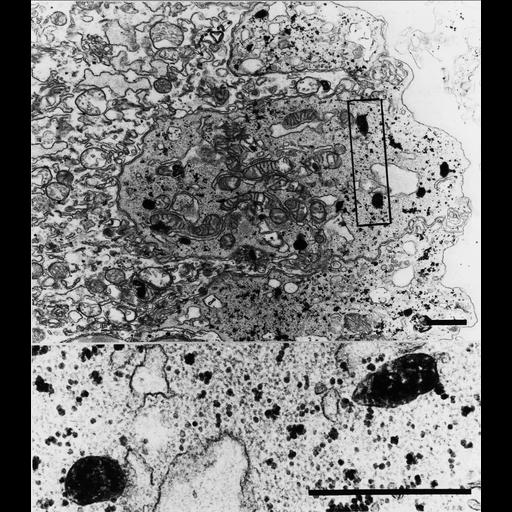
Many oval peroxisomes can be seen in normal cytotrophoblast cells of first trimester chorionic villus. The cytotrophoblast also contains glycogen particles, isolated and in flakes, which distinguish these cells from the syncytiotrophoblast (at left in this image). Bars = 1 µm (low magnification), 0.1 µm (high magnification). In some genetic diseases (Zellweger, Infantile Refsum) peroxisomes are absent; this can be diagnosed in chorionic villi.

Technical Details

Peroxisomes are contrasted with DAB at pH 10.5 by their catalase activity, after fixation in buffered formal-calcium; post-fixed in OSO4

Biological Sources
NCBI Organism Classification
Homo sapiens
Cell Type
mononuclear cytotrophoblast cell
Cellular Component
peroxisome
Attribution
Names
Frank Roels
Published
"Peroxisomes. A personal account". pp. 151. Copyright VUB-Press 1991.
Citation
Digital Object Identifier (DOI)
doi:10.7295/W9CIL46754
Archival Resource Key (ARK)
ark:/b7295/w9cil46754