Alternate header for print version


Licensing
Attribution Non-Commercial Share Alike:This image is licensed under a Creative Commons Attribution, Non-Commercial Share Alike License. View License Deed | View Legal Code
tweet  
*CIL – Cell Image Library accession number. Please use this to reference an image.

CIL:47808*  Cite 
Description

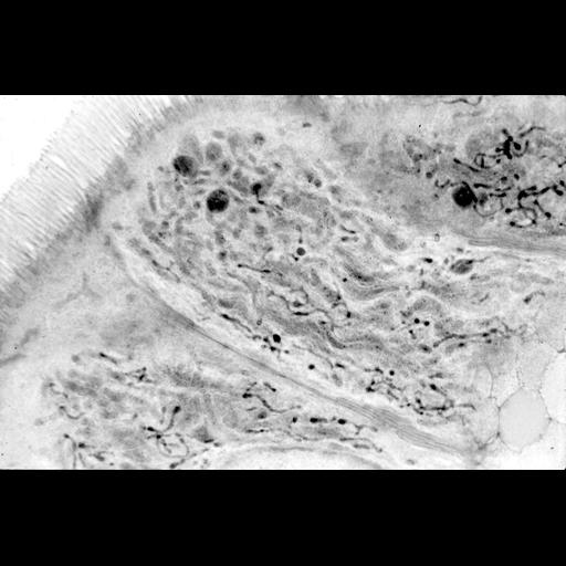
Human duodenal epithelium. Peroxisomes are stained with DAB at pH 10.5 by their catalase activity and postosmicated. 0.5 µm plastic section were visualized using electron microscopy at low magnification. Image shows thin worm like peroxisomes. Three large dark round structures are lysosomes. Brush border microvilli are well seen. More details in Roels et al: http://gut.bmj.com/content/32/8/858.long

Biological Sources
NCBI Organism Classification
Homo sapiens
Cell Type
duct epithelial cell
Cellular Component
peroxisome
Biological Context
Molecular Function
catalase activity
Attribution
Names
Frank Roels
Marina Pauwels
Peter Van der Spek
Citation
Digital Object Identifier (DOI)
doi:10.7295/W9CIL47808
Archival Resource Key (ARK)
ark:/b7295/w9cil47808