Images are acquired from fixed and living heart tissue using fiber-optics and laser-scanning confocal microscopy, respectively. Three sets of images denoted as CCM, FCMtopical, FCMcarrier were stored for subsequent analyses.
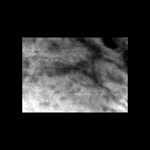
Tissue regions were labeled with a dye carrier affixed to the tip of a fiber-optics microprobe (UltraMiniOWD30; Mauna Kea Technologies). Dye carriers were loaded for 15 min with sodium fluorescein [Fluorescite® (fluorescein injection, USP) 10%; Alcon, Fort Worth, TX, US; 1:1000] prior to imaging. With this imaging protocol, we acquired two-dimensional image sequences of the tissue regions at a lateral resolution, optical sectioning, field of view, frame rate, and z-scan range of 1.4 μm, 7 μm, 186 by 130 μm, 12 Hz and 26 μm, respectively. Example images from these image sequences were stored as FCMcarrier.
| Spatial Axis | Image Size | Pixel Size |
|---|---|---|
| X | —— | 186µm |
| Y | —— | 130µm |
| Z | —— | 26µm |