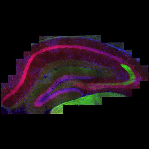
High-resolution, large-scale mosaic image, for mouse brain coronal section through the dorsal hippocampus, identified distinct localization of PKA RIβ and RIIβ regulatory subunits across various hippocampal subfields. Triple labeled for Nuclei (Hoescht stain, blue), RIβ (cy3, red) and RIIβ (Alexa 488, green). This image is a maximum projection of the Z-stack through the tissue, and has been downsampled from the raw data image, which can be accessed using the link provided to the Cell Image Library.
Mouse brain was perfused with 4% paraformaldehyde, sectioned through the hippocampus in the coronal plane on a vibratome (100 µm). The section was immunostained with RIβ and RIIβ primary antibodies. Secondary antibodies: Alexa Fluor 488 Donkey anti rabbit (green) and CY3 Donkey anti sheep (red). Images were acquired with a FluoView 1000 (Olympus Center Valley, PA,USA) using 60X objective and a high-precision motorized stage to collect the large scale mosaics. The specimen was excited sequentially with a laser at two different wavelengths: 488nm and 561nm. The boundaries (in x, y, and z) of the tissue section were defined using the Multi-Area Time Lapse function of ASW 1.7c microscope operating-software provided by Olympus (Olympus, Center Valley, PA, USA). The software automatically generates a list of 3D stage positions covering the volume of interest, which are computed using the dimensions of a single image in microns, degree of overlap between adjacent images and z-step size. Overlap between two adjacent images (x-y) was 10% and the z-step was ~0.5 mm/section; there is no overlap in z. image normalization was performed using ImageJ. Reconstruction Data File Montage of a mouse coronal section cut through dorsal hippocampus, triple labeled for Hoescht stain (blue), PKA RIβ regulatory subunit (red), and PKA RIIβ regulatory subunit (green). Reconstruction Data Files, mosaic reconstructed with 10% overlap; image normalization performed using imageJ.
| Spatial Axis | Image Size | Pixel Size |
|---|---|---|
| X | 13894px | —— |
| X | 7447px | —— |