Alternate header for print version


Licensing
Attribution Non-Commercial Share Alike:This image is licensed under a Creative Commons Attribution, Non-Commercial Share Alike License. View License Deed | View Legal Code
tweet  
*CIL – Cell Image Library accession number. Please use this to reference an image.

CIL:50638*  Cite 
Description

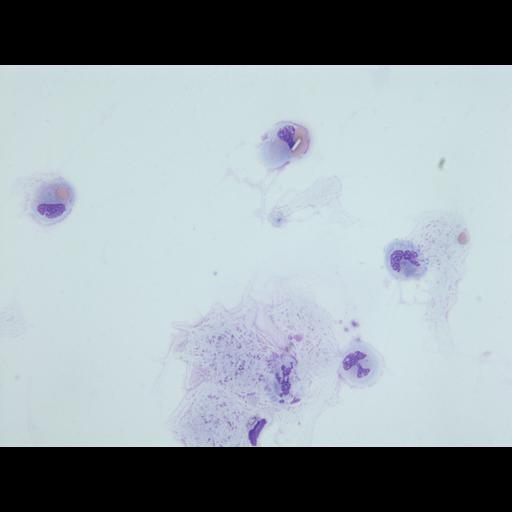
Two monocyte-type cells showed erythrocytes either at contact or intruding in the monocyte cytoplasm, For one of the cells, the erythrocyte was covered with cytoplams-like material. For the other monocyte, a rectangular crystal could be identifiedat contact to the monocyte nucleus and superposed possibly to the erythrocyte. Other monocyte/polymorphonuclear type cells showing nuclei with aberant shapes, in part lytic, could be observed.

Technical Details

Cerebrospinal fluid, cytocentrifugation, May-Grunwald-Giemsa stain.

Biological Sources
NCBI Organism Classification
Homo sapiens
Cell Type
monocyte
erythrocyte
Cellular Component
Cytoplasm lesion
Biological Context
Biological Process
Erytrophagocytosis
Attribution
Names
Adriana Handra-Luca,
APHP University Paris Nord
Citation
Digital Object Identifier (DOI)
doi:10.7295/W9CIL50638
Archival Resource Key (ARK)
ark:/b7295/w9cil50638
Imaging
Image Mode
microscopy
Visualization Methods
x100