Alternate header for print version


Licensing
Attribution Only: This image is licensed under a Creative Commons Attribution License. View License Deed | View Legal Code
tweet  
*CIL – Cell Image Library accession number. Please use this to reference an image.

CIL:50721*  Cite 
Description

FIB-SEM Dataset of anti-PKHD1L1 Immuno-Gold Labeled Outer Hair Cell Stereocilia Bundles. Postnatal day 4 mouse Inner Ear Organ of Corti labeled with anti-PKHD1L1 antibody (10 nm gold beads). The dataset represents the apical end of the Outer Hair Cell with the stereocilium bundle. Images were acquired with FEI Helios FIB-SEM, using backscatter detector from an epoxy resin embedded inner ear Organ of Corti sample.

Technical Details

Images were acquired with FEI Helios FIB-SEM, using backscatter detector from an epoxy resin embedded inner ear Organ of Corti sample.

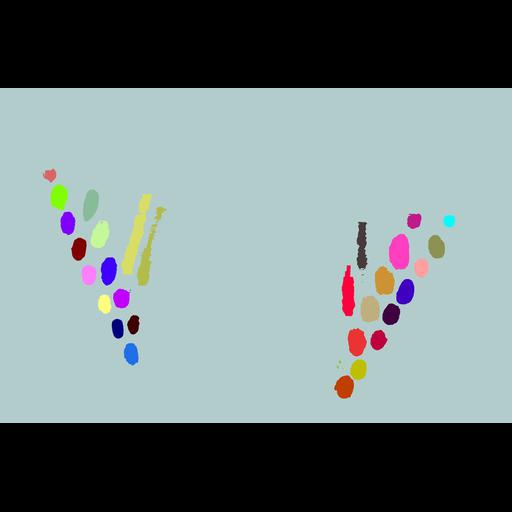
6.3_Cell_6_Stereocilia_Segmentation is a multipage TIFF file illustrating stereocilia segmentation results. Same as ‘Towers.am’ file used for MATLAB volume analysis of this cell, but converted to TIFF to allow for its online preview. File contains 324 images, as all the planes that contained no segmentation data were removed.

This file is a part of Dataset 6, comprised of files related to the Cell #6 of current study. The numbering order of the files reflects the order of operations performed during the analysis. The raw imaging data related to this cell was deposited within Dataset 1 (CIL:50680).

Biological Sources
NCBI Organism Classification
Mus musculus
Cell Type
Inner Ear Outer Hair Cell
Cellular Component
Hair Cell stereocilia
Cuticular plate
Mitochondria
Polycystic Kidney and Hepatic Disease 1-Like 1 (PKHD1L1) labeling (gold beads)
Biological Context
Biological Process
Native auditory Hair Cells at postnatal day 4
Attribution
Names
Maryna V. Ivanchenko
Marcelo Cicconet
Hoor Al Jandal
Xudong Wu
David P. Corey (http://corey.med.harvard.edu/)
Artur A. Indzhykulian (https://ailab.hms.harvard.edu/)
Link
Scientific Data article
Nature Communications article
OTHER
Massachusetts Eye and Ear; Harvard Medical School
6.3_Cell_6_Stereocilia_Segmentation
Citation
Digital Object Identifier (DOI)
doi:10.7295/W9CIL50721
Archival Resource Key (ARK)
ark:/b7295/w9cil50721
Grouping This image is part of a group.
Imaging
Image Mode
Serial Electron Microscopy
Focused Ion Beam Scanning Electron Microscopy (FIB-SEM)
Visualization Methods
Heavy metal stain (Osmium Tetroxide, Potassium Ferrocyanide, Tannic Acid)
Immunogold labeling with 10 nm gold beads