Alternate header for print version


Licensing
Attribution Only: This image is licensed under a Creative Commons Attribution License. View License Deed | View Legal Code
tweet  
*CIL – Cell Image Library accession number. Please use this to reference an image.

CIL:50727*  Cite 
Description

FIB-SEM Dataset of anti-PKHD1L1 Immuno-Gold Labeled Outer Hair Cell Stereocilia Bundles. Postnatal day 4 mouse Inner Ear Organ of Corti labeled with anti-PKHD1L1 antibody (10 nm gold beads). The dataset represents the apical end of the Outer Hair Cell with the stereocilium bundle. Images were acquired with FEI Helios FIB-SEM, using backscatter detector from an epoxy resin embedded inner ear Organ of Corti sample.

Technical Details

Images were acquired with FEI Helios FIB-SEM, using backscatter detector from an epoxy resin embedded inner ear Organ of Corti sample.

7.3_Test_Cell_MATLAB_Volume_Analysis_Results is a compressed (zipped) folder containing all files required to execute the MATLAB volume analysis. The folder also includes the resulting output files and figures. 
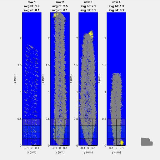
This file is a part of Dataset 7, comprised of files used as a technical validation dataset. It is an artificial dataset with manually placed gold beads, used to access the accuracy of the MATLAB script. The choice of gold bead placement location was unique for each row of stereocilia to control for potential errors of stereocilia placement within the rows during MATLAB analysis. No beads were placed on the surface of the kinocilium.

Biological Sources
NCBI Organism Classification
Mus musculus
Cell Type
Inner Ear Outer Hair Cell
Cellular Component
Hair Cell stereocilia
Cuticular plate
Mitochondria
Polycystic Kidney and Hepatic Disease 1-Like 1 (PKHD1L1) labeling (gold beads)
Biological Context
Biological Process
Native auditory Hair Cells at postnatal day 4
Attribution
Names
Maryna V. Ivanchenko
Marcelo Cicconet
Hoor Al Jandal
Xudong Wu
David P. Corey (http://corey.med.harvard.edu/)
Artur A. Indzhykulian (https://ailab.hms.harvard.edu/)
Link
Scientific Data article
Nature Communications article
OTHER
Massachusetts Eye and Ear; Harvard Medical School
7.3_Test_Cell_MATLAB_Volume_Analysis_Results
Citation
Digital Object Identifier (DOI)
doi:10.7295/W9CIL50727
Archival Resource Key (ARK)
ark:/b7295/w9cil50727
Grouping This image is part of a group.
Imaging
Image Mode
Serial Electron Microscopy
Focused Ion Beam Scanning Electron Microscopy (FIB-SEM)
Visualization Methods
Heavy metal stain (Osmium Tetroxide, Potassium Ferrocyanide, Tannic Acid)
Immunogold labeling with 10 nm gold beads