Alternate header for print version


Licensing
Attribution Non-Commercial Share Alike:This image is licensed under a Creative Commons Attribution, Non-Commercial Share Alike License. View License Deed | View Legal Code
tweet  
*CIL – Cell Image Library accession number. Please use this to reference an image.

CIL:52464*  Cite 
Description

Tile x=1, y=5, z=19 from BL32077_Myc_Terc

Technical Details

One z layer of a z-stack at the given tile position

Biological Sources
NCBI Organism Classification
Homo sapiens
Cell Type
High-grade serious overian cancer
Cellular Component
Copy number of the hTERT, c-MYC, and SE7 gene regions determined by colored probes
Biological Context
Biological Process
DNA (stained by DAPI) as well as the hTERT, c-MYC, and SE7 gene regions (marked by coloured probes)
Attribution
Names
Anna Piskorz
Link
Cancer Research UK Cambridge Institute
OTHER
W9P20452
Citation
Digital Object Identifier (DOI)
doi:10.7295/W9CIL52464
Archival Resource Key (ARK)
ark:/b7295/w9cil52464
Grouping This image is part of a group.
Imaging
Image Mode
Fluorescence in situ hybridization (FISH)
Visualization Methods
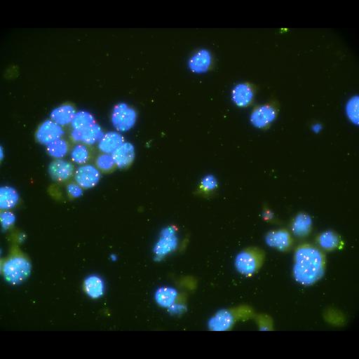
Nikon Eclipse fluorescence inverted microscope equipped with a charge-coupled device camera (Andor Neo sCMOS), using filter sets for DAPI/ YGFP/TRITC/CY GFP with an objective lens (Plan Apo VC 100x, Nikon). Captured with 100x magnification of the objective and a pixel size of 0.07 microns