Alternate header for print version


Licensing
Attribution Non-Commercial Share Alike:This image is licensed under a Creative Commons Attribution, Non-Commercial Share Alike License. View License Deed | View Legal Code
tweet  
*CIL – Cell Image Library accession number. Please use this to reference an image.

CIL:54644*  Cite 
Description

We examined the structural organization of cytoskeletal components and membrane systems in neurons of well preserved biopsy material from Alzheimer’s disease (AD). Information was obtained from thin sectioned material using conventional electron microscopy and from thick sections with the high voltage electron microscope.

Technical Details

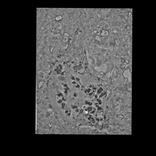
Degenerating neuron with PHF and lipofuscin. Extremely degraded with laterally displaced nucleus.

Biological Sources
NCBI Organism Classification
Homo sapiens
Cell Type
pyramidal cell
neuron of cerebral cortex
Cellular Component
neuronal cell body
Biological Context
Human Disease
Alzheimer's disease
Attribution
Names
Mark Ellisman
Tom Deerinck
Matthew Madany
OTHER
Negative 3
Citation
Digital Object Identifier (DOI)
doi:10.7295/W9CIL54644
Archival Resource Key (ARK)
ark:/b7295/w9cil54644
Grouping This image is part of a group.
Imaging
Image Type
Serial section
Image Mode
transmission electron microscopy (TEM)