Using a camera mounted above the microscope, experts adjust the lens and angle to capture the stain and transfer the image to a computer for processing.
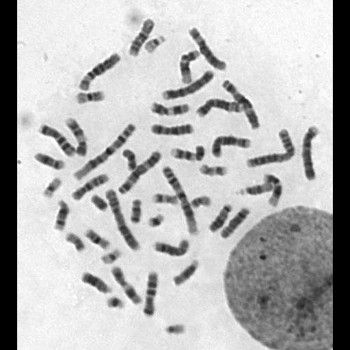
We use in-situ harvest method and trypsin/Wright stain procedure to prepare G-banding. Briefly, the amniotic cells were cultured in BIO-AMF medium (Biological industrial, Beit-Haemek, Israel) for 6 to 8 days.Treating cells with colcemid (NY, USA, Gibco) for 30 min to arrest cells at metaphase. Swell cells with hypotonic solution and fix with methanol/acetic acid mixture. Fixed cells were treated with tryspin then stained with Wright stain solution (MO, Sigma-Aldrich). The karyotype was interpreted according to The International System for Human Cytogenomic Nomenclature (ISCN).
See also: M. J. Barch, T. Kuntsen and J. L. Spurbeck, The AGT Cytogenetics Laboratory Manual, Lippincott-Raven (1997). Yunis J. High resolution of human chromosome. Science 1976: 191:1268-1270. J. McGowan-Jordan, A.
Sim+A13ons and M. Schmid. An International System for Human Cytogenetic Nomenclature. KARGER (2016).