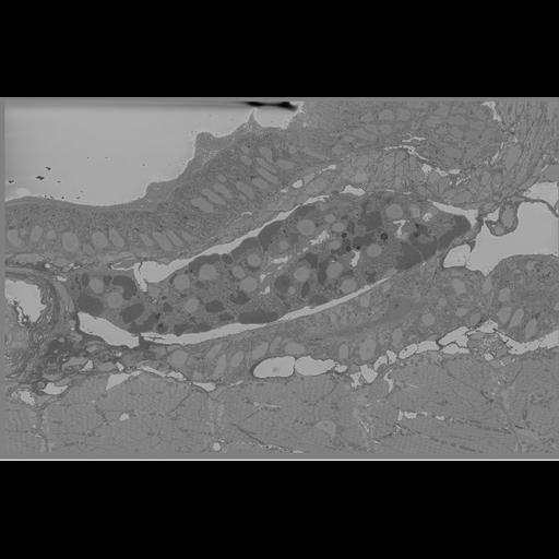
Sagittal sections of 5 days post-fertilization cd41:gfp+ zebrafish larva in the region of the anterior kidney (eLife Table 1: cd41:GFP 5 dpf)
Larvae preparation for GFP+ CLEM (eLife: Workflow #2). To facilitate the 3D correlation between different imaging modalities when using any GFP+ transgenic line, we added DRAQ5, a nuclear DNA binding fluorescent dye. Briefly, the 5 dpf transgenic zebrafish larvae were fixed with 0.5% glutaraldehyde and 2% paraformaldehyde in 0.15M cacodylate buffer (CB, pH 7.4) for 2.5 hours and 100 m thick sagittal sections were collected and incubated in DRAQ5 (1:1000, Cell Signaling Technology) on ice for an hour. Confocal images of GFP and DRAQ5 signals were collected on a Leica SPE II confocal microscope with a 20X oil-immersion objective lens using 488 nm and 633 nm excitation. Following, the samples were preincubated in 2.5 mM DAB solution (25.24 mM stock in 0.1 M HCl) in 0.15 M CB for 30 min on ice. Next, the photo-oxidation of DAB by DRAQ5 was done in 2.5 mM DAB in 0.15 M CB using a solar simulator (Spectra-Physics 92191–1000 solar simulator with 1600 W mercury arc lamp and two Spectra-Physics SP66239–3767 dichroic mirrors to remove infrared and ultraviolet wavelengths), while bubbling oxygen in the solution. The light was filtered through a 10 cm square bandpass filters (Chroma Technology Corp.) for illumination at 615 nm (40 nm band pass). The reaction was monitored every 20 minutes and stopped when the desired darkening in the nuclei was achieved. Larvae were then washed 5 times with 0.15 M CB and incubated in 2% OsO4/1.5% potassium ferrocyanide in 0.15 M CB containing 2 mM CaCl2 to get an EM visible stain. The larvae were left for 30 min on ice and then 30 min at room temperature (RT). After thorough washing in double distilled water (ddH2O), larvae were placed into 0.05% thiocarbohydrazide for 30 min. Larvae were again washed and then stained with 2% aqueous OsO4 for 30 min. Larvae were washed and then placed into 2% aqueous uranyl acetate overnight at 4°C. Larvae were washed with ddH2O at RT and then stained with 0.05% en-bloc lead aspartate for 30 min at 60°C. Larvae were washed with ddH2O and then dehydrated on ice in 50%, 70%, 90%, 100%, 100% ethanol solutions for 10 min at each step. Larvae were then washed twice with dry acetone and placed into 50:50 Durcupan ACM:acetone overnight. Larvae were transferred to 100% Durcupan resin overnight. Larvae were then flat embedded between glass slides coated with mould-release compound and left in an oven at 60°C for 72 h. The larva block was imaged with a Merlin scanning electron microscope equipped with a 3View2XP and OnPoint backscatter detector. The volume was collected at 3 kV, with 10.8 nm pixels x and y ; 70 nm Z steps and 70% local gas injection. The raster size was 25k x 12k and the volume was aligned using cross correlation and visualized using IMOD.