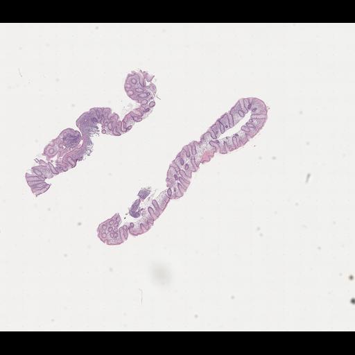
Scan 10_H_CA_D -- The dataset consists of 951 tissue section scans taken from 167 H&E-stained whole slides from 18 pediatric inflammatory bowel disease (IBD) patients. Slide section scans are provided as-is without any initial image pre-processing, normalization, or cropping. Labels are provided at the patient, slide (anatomic site), and section (tissue slice) levels. Each slide is labelled with biopsy site and normal or abnormal classification per the surgical pathology report. Each tissue section scan from an abnormal slide is further classified as normal or abnormal by pathologist. Files may be viewed and annotated using standard, opensource slide-viewing software, such as Aperio ImageScope, OpenSlide, TiffSlide, and QuPath.
Scans of H&E-stained histology slides of biopsied tissue from endoscopy. Captured with MikroScan SL5 in SVS TIFF format with .tif file extension.