Alternate header for print version


Licensing
Public Domain: This image is in the public domain and thus free of any copyright restrictions. However, as is the norm in scientific publishing and as a matter of courtesy, any user should credit the content provider for any public or private use of this image whenever possible. Learn more
tweet  
*CIL – Cell Image Library accession number. Please use this to reference an image.

CIL:6517*  Cite 
Description

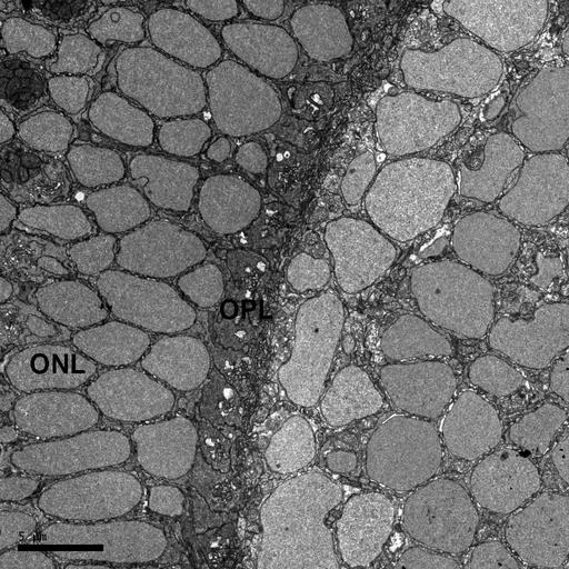
This study of the zebrafish eye is a series of four images taken at increasing magnifications. The first image is the SEM block face of the zebrafish eye. This is followed by this image, the second image, which is a higher magnification TEM image of the plexiform layer and the outer nuclear layer of the zebrafish retina. The third image is a 15,000X TEM image of the bipolar and horizontal cells interdigitating in the OPL layer of the zebrafish retina. The fourth image in the series is a higher magnification (60,000X) of the dense synaptic bar and synaptic vesicles. TEM image of larval zebrafish retina showing outer nuclear layer (ONL) and outer plexiform layer (OPL). Image collected on a JOEL 1230 at 80 kV using a Gatan 967 slow-scan, cooled CCD camera. 70 nm thick section. Microwave processing, osmium tetroxoxide/osmid procedure for enhancing membranes, and copper lead en bloc staining. Procedure for specimen preparation available at http://www.stanford.edu/~redhair/JoAnn_Buchanan.

Biological Sources
NCBI Organism Classification
Danio rerio
Cell Type
Outer plexiform layer of retina
Outer nuclear layer of retina
Biological Context
Biological Process
retina layer formation
Zebrafish Anatomy
retina
retinal outer nuclear layer
retinal outer plexiform layer
Attribution
Names
JoAnn Buchanan (Stanford)
Link
http://www.stanford.edu/~redhair/JoAnn_Buchan...
Citation
Digital Object Identifier (DOI)
doi:10.7295/W9CIL6517
Archival Resource Key (ARK)
ark:/b7295/w9cil6517
Grouping This image is part of a group.
Imaging
Image Type
recorded image
Image Mode
transmission electron microscopy (TEM)
Parameters Imaged
electron density
Source of Contrast
differences in adsorption or binding of stain
Visualization Methods
osmium tetroxide
lead salt
copper salt
Processing History
unprocessed raw data
Dimensions
Spatial Axis Image Size Pixel Size
X 1024px 0.032µm
Y 1024px 0.032µm