Alternate header for print version


Licensing
Public Domain: This image is in the public domain and thus free of any copyright restrictions. However, as is the norm in scientific publishing and as a matter of courtesy, any user should credit the content provider for any public or private use of this image whenever possible. Learn more
tweet  
*CIL – Cell Image Library accession number. Please use this to reference an image.

CIL:7331*  Cite 
Description

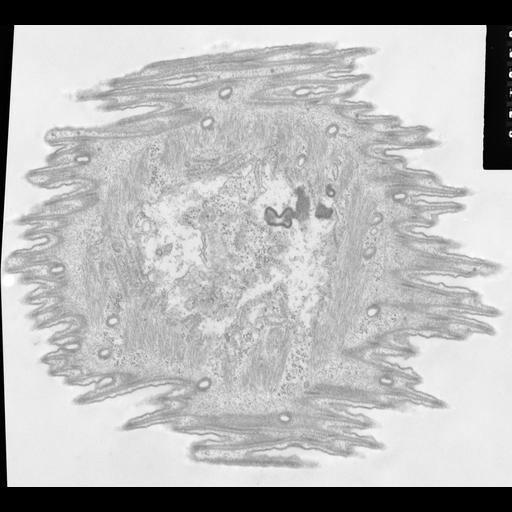
High resolution of the layer of contractile myonemes in Opercularia coarctata that covers most of the cytosolic side of the pellicle. Pellicular pores (the clathrin coated pits of ciliates) penetrate through this sheet. The ER is spread out against this sheet and, besides having decorated tubules, it bears thinner rails that lie between the ER and the myonemes and spindle-shaped mid pieces. For a description of these rails and mid pieces as they are found in peritrichs. Their function still remains to be determined. TEM taken on 6/7/69 by R. Allen with Philips 300 operating at 60kV. Neg. 14,800X. The raw film was scanned with an Epson Perfection V750 Pro. this image is suitable for quantitative analysis. Standard glutaraldehyde fixation followed by osmium tetroxide, dehydrated in alcohol and embedded in an epoxy resin. Microtome sections prepared at approximately 75nm thickness. Additional information available at (http://www5.pbrc.hawaii.edu/allen/).

Biological Sources
NCBI Organism Classification
Opercularia [NCBITaxon:168247]
Opercularia coarctata
Cell Type
cell by organism
eukaryotic cell
Eukaryotic Protist
Ciliated Protist
Cell Line
Cambridge Culture Collection, Cambridge, England, # LB1657/1
Cellular Component
contractile myonemes
smooth endoplasmic reticulum
clathrin coat of coated pit
Biological Context
Biological Process
contractile fiber organization
membrane corridors
Molecular Function
structural constituent of cytoskeleton
GO:0015570
Attribution
Names
Richard Allen
Citation
Digital Object Identifier (DOI)
doi:10.7295/W9CIL7331
Archival Resource Key (ARK)
ark:/b7295/w9cil7331
Grouping This image is part of a group.
Imaging
Image Type
transmission electron microscopy (TEM)
illumination by electrons
Image Mode
detection of electrons
film
Parameters Imaged
electron density
Source of Contrast
stain with broad specificity
Visualization Methods
stain with broad specificity
osmium tetroxide
uranyl salt
lead salt
Processing History
recorded image
film
unprocessed raw data
Data Qualifiers
raw, unprocessed data
suitable for spatial measurements
Dimensions
Spatial Axis Image Size Pixel Size
X 5040px 1nm
Y 4548px 1nm