Alternate header for print version


Licensing
Public Domain: This image is in the public domain and thus free of any copyright restrictions. However, as is the norm in scientific publishing and as a matter of courtesy, any user should credit the content provider for any public or private use of this image whenever possible. Learn more
tweet  
*CIL – Cell Image Library accession number. Please use this to reference an image.

CIL:808*  Cite 
Description

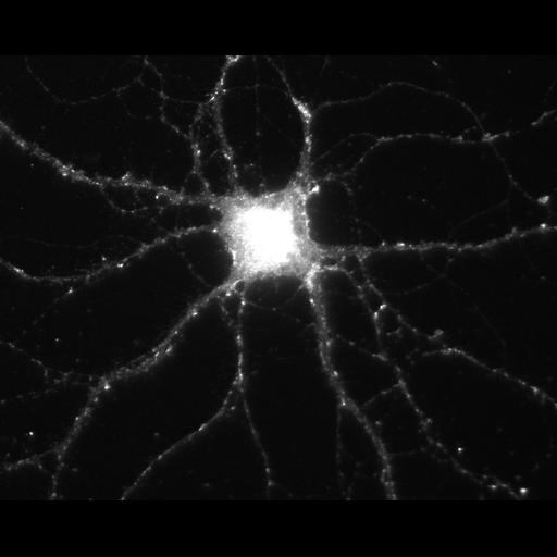
During hippocampal neuron development in vitro, N-cadherin becomes selectively localized to excitatory synapses. This image shows immunolocalization of the NMDA receptor, which is postsynaptic. Other images in this image group show immunolocalization of N-cadherin, colocalized at synapses that are also immunopositive for the presynaptic glutamate transporters V-glut 1 and 2. Individual fluorescent images were acquired using a Nikon Diaphot 300 with a 60X objective. Preparation was fixed in 4% paraformaldehyde and permeabilized with .01% TritonX-100.

Biological Sources
NCBI Organism Classification
Rattus
Cell Type
multipolar neuron
Cellular Component
excitatory synapse
synapse part
Attribution
Names
Tonya Anderson
Deanna Benson
Citation
Digital Object Identifier (DOI)
doi:10.7295/W9CIL808
Archival Resource Key (ARK)
ark:/b7295/w9cil808
Grouping This image is part of a group.
Imaging
Image Type
recorded image
Image Mode
fluorescence microscopy
Parameters Imaged
fluorescence emission
Source of Contrast
distribution of epitope
Visualization Methods
primary antibody plus labeled secondary antibody
Processing History
unprocessed raw data
Sample Preparation
Methods
formaldehyde fixed tissue
detergent permeabilized
Relation To Intact Cell
dispersed cells in vitro
Dimensions
Spatial Axis Image Size Pixel Size
X 1280px 0.113µm
Y 1024px 0.113µm