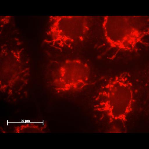
This culture of HeLa cells was grown, fixed and labeled directly on 35-mm plastic petri dishes (not on cover slips). To preserve the 3D structure of cells, cultures were fixed in formaldehyde vapors at room temperature over 20 min. Fixation was performed as follows: medium was completely aspirated, a 50ul drop of 37% formaldehyde was placed on the lid of the dish and covered with the dish of cells, bottom up. After fixation dishes were carefully filled with 1ml of 50% glycerol in PBS and dishes were stored at -20 C until staining. Before staining, cells were incubated with blocking solution (5% fetal calf serum, PBS, 0.2% triton x-100, 25 min room temp.), then treated with primary antibody, which was mouse anti-Cytochrome C from BD Pharmigen (cat.# 556433) diluted 1:100 in blocking solution, 1h room temp. After standard washing (PBS 3-4 times, 5 min each), secondary antibody (donkey monovalent Fab-fragment anti-mouse IgG conjugated with Rhodamine Red-X from Jackson ImmunoResearch laboratories, concentration 2.5ug/ml) was applied. Images were captured on a Zeiss Axioplan-2. Objective - 63x oil immersion/NA 1.25. Magnifying lens (optovar) was 1.5. Digital camera – Hamamatsu ORCA-ER. Program – AxioVision 3.5.1. Illumination-mercury bulb HBP50. Filter set – standard for Rhodamine/Cy3/Alexa 564 – excitation 530-585 and emission LP 615. Mounting medium-VectaShield.
| Spatial Axis | Image Size | Pixel Size |
|---|---|---|
| X | 2560px | 32.69nm |
| Y | 2016px | 32.69nm |