Alternate header for print version


Licensing
Public Domain: This image is in the public domain and thus free of any copyright restrictions. However, as is the norm in scientific publishing and as a matter of courtesy, any user should credit the content provider for any public or private use of this image whenever possible. Learn more
tweet  
*CIL – Cell Image Library accession number. Please use this to reference an image.

CIL:9015*  Cite 
Description

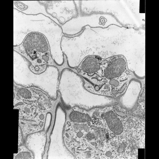
A high resolution micrograph of the monokinetids (single somatic basal bodies) of Coleps hirtus bear an anteriorly directed striated fibrous kinetodesmal fiber and a posteriorly directed ribbon of postciliary microtubules that arise from the posterior side of each basal body. Both pass to the same side of the row of basal bodies. Transverse microtubules arise from the anterior side of the basal body and pass to the other side of the row of basal bodies. These structures presumably give support to the twisting movements of the beating cilium. Standard glutaraldehyde fixation followed by osmium tetroxide, dehydrated in alcohol and embedded in an epoxy resin. Microtome sections prepared at approximately 75nm thickness. TEM taken on 5/23/69 by R. Allen with Philips 300 operating at 60kV. Neg. 14,800X. The raw film was scanned with an Epson Perfection V750 Pro. This high resolution image is suitable for quantitative analysis Additional information available at (http://www5.pbrc.hawaii.edu/allen/).

Biological Sources
NCBI Organism Classification
Coleps hirtus
Cell Type
eukaryotic cell
Eukaryotic Protist
Ciliated Protist
Cell Line
Cambridge Culture Collection, Cambridge, England, catalog # LB1656/1
Cellular Component
cilium axoneme
microtubule basal body
axonemal microtubule
plasma membrane
cytoskeleton
microtubule cytoskeleton
actin cytoskeleton
striated basal body fiber
associated microtubules
Biological Context
Biological Process
ciliary or flagellar motility
microtubule cytoskeleton organization
microfibril organization
Molecular Function
structural molecule activity
Attribution
Names
Richard Allen
Citation
Digital Object Identifier (DOI)
doi:10.7295/W9CIL9015
Archival Resource Key (ARK)
ark:/b7295/w9cil9015
Grouping This image is part of a group.
Imaging
Image Type
transmission electron microscopy (TEM)
illumination by electrons
Image Mode
detection of electrons
film
Parameters Imaged
electron density
Source of Contrast
stain with broad specificity
Visualization Methods
stain with broad specificity
osmium tetroxide
uranyl salt
lead salt
Processing History
recorded image
film
unprocessed raw data
Data Qualifiers
raw, unprocessed data
suitable for spatial measurements
Dimensions
Spatial Axis Image Size Pixel Size
X 5152px 1nm
Y 6184px 1nm