Alternate header for print version


Licensing
Public Domain: This image is in the public domain and thus free of any copyright restrictions. However, as is the norm in scientific publishing and as a matter of courtesy, any user should credit the content provider for any public or private use of this image whenever possible. Learn more
tweet  
*CIL – Cell Image Library accession number. Please use this to reference an image.

CIL:9525*  Cite 
Description

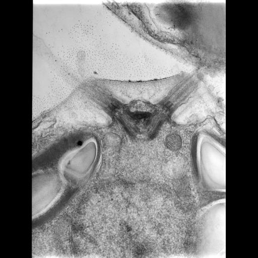
The unicellular alga Chlamydomonas reinhardtii has a pair of anterior flagella that function in motility. In this image, the base of the flagella with basal bodies and associated structures are are cut longitudinally. Below is the spherical nucleus and portions of chloroplast.

Technical Details

250 nm thick epon sections were recorded with a high voltage JEM-1000 TEM at 1000KV.

Biological Sources
NCBI Organism Classification
Chlamydomonas reinhardtii
Cell Type
plant cell
Cell Line
strain in culture
Cellular Component
chloroplast
nucleus
microtubule basal body
flagellum part
Biological Context
Biological Process
photosynthesis, light harvesting
organelle organization
Attribution
Names
Mary Morphew
J. Richard McIntosh
Citation
Digital Object Identifier (DOI)
doi:10.7295/W9CIL9525
Archival Resource Key (ARK)
ark:/b7295/w9cil9525
Grouping This image is part of a group.
Imaging
Image Type
transmission electron microscopy (TEM)
film
Image Mode
FBbi:00000622
detection of electrons
Parameters Imaged
electron density
Source of Contrast
elastic scattering of electrons
Visualization Methods
uranyl salt
Processing History
unprocessed raw data
Sample Preparation
Methods
glutaraldehyde fixed tissue
tissue in plastic embedment
Relation To Intact Cell
microtome-sectioned tissue
Dimensions
Spatial Axis Image Size Pixel Size
X 3447px 0.8nm
Y 4580px 0.8nm