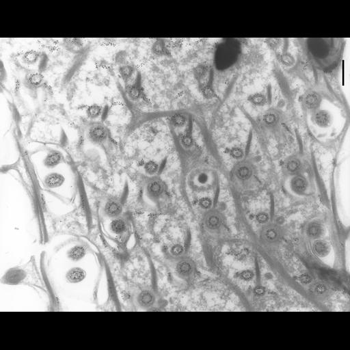
Surface pattern of P. tetraurelia in a region with two basal bodies in each cortical unit. This is a thin-frozen section labeled with immunogold showing the location of b-tubulin. The gold lies over the microtubules of the x-sectioned basal bodies and ciliary axonemes. It also labels tangentially sectioned transverse and postciliary microtubules. Neither the infraciliary lattice, the less obvious striated bands or the kinetodesmal fibers are labeled with anti-b-tubulin. TEM taken on 7/8/96 by R. Allen with Zeiss 10A operating at 80kV. Neg. 12,000X. Bar = 0.5µm. Cells were lightly fixed with 0.25% glutaraldehyde and infiltrated with 2.3M sucrose before being frozen in liquid nitrogen and thin sectioned at a temperature of –100°C at approximately 75nm thickness. Frozen sections from these preparations were then thawed, washed, and exposed to a monoclonal primary antibody that was raised in mice or rabbit/goat and to colloidal gold-complexed goat-anti-mouse/rabbit secondary antibodies. Further details of preparation are detailed in Methods Cell Biol. 2010;96:143-73. The negative was printed to paper and the image was scanned to Photoshop. This digitized image is available for qualitative analysis. An unprocessed, high resolution version of this image (CIL:1312) is in the library and available for quantitative analysis. Additional information available at (http://www5.pbrc.hawaii.edu/allen/).
| Spatial Axis | Image Size | Pixel Size |
|---|---|---|
| X | 2734px | —— |
| Y | 2146px | —— |