Alternate header for print version


Licensing
Public Domain: This image is in the public domain and thus free of any copyright restrictions. However, as is the norm in scientific publishing and as a matter of courtesy, any user should credit the content provider for any public or private use of this image whenever possible. Learn more
tweet  
*CIL – Cell Image Library accession number. Please use this to reference an image.

CIL:9975*  Cite 
Description

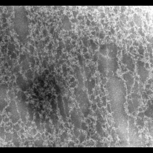
Newt (Notopthalmus viridescens erythrocytes were isolated in 0.1M KCl, spread on the surface of Na-citrate, picked up on formvar-carbon films, fixed with glutaraldehyde and paraformaldehyde and negatively stained with 2% sodium phosphotungstate. Images were recorded at 40KX and accelerating voltage of 1MeV at the University of Wisconsin HVEM facility.This image was taken with a specimen tilt of 45 degrees. A grouped image of the same field at a 55 degree tilt provides an oblique stereo view of the tangle of chromatin fibers in the dispersed nucleus.

Biological Sources
NCBI Organism Classification
notopthalmus viridescens
Cell Type
erythrocyte
Cellular Component
nuclear chromatin
Biological Context
Biological Process
DNA packaging
Attribution
Names
Hans Ris
Pubmed
7022124
Citation
Digital Object Identifier (DOI)
doi:10.7295/W9CIL9975
Archival Resource Key (ARK)
ark:/b7295/w9cil9975
Grouping This image is part of a group.
Sample Preparation
Methods
glutaraldehyde fixed tissue
formaldehyde fixed tissue
Relation To Intact Cell
spread nucleus
Dimensions
Spatial Axis Image Size Pixel Size
X 2956px 0.5nm
Y 2742px 0.5nm