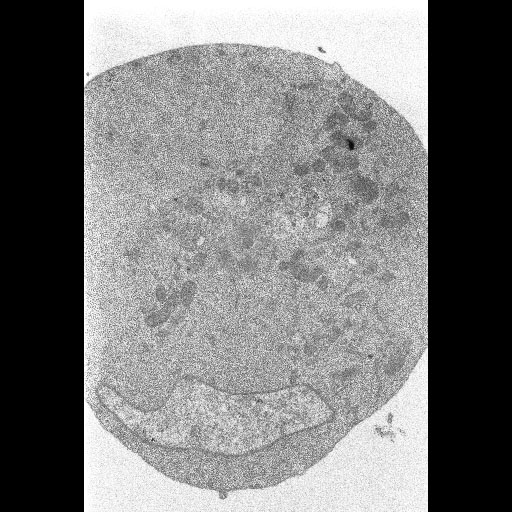
Electron micrograph of a cultured Drosophila DL1 cell infected with flock house virus, prepared using chemical fixation followed by routine embedding for electron microscopy. This specimen was prepared as a control to compare different high pressure freezing protocols.
Full resolution image description
Full sized tiff image (CAF_rec.tif) of the insect cells processed using conventional aldehyde fixation and routine embedding for electron microscopy. Image corresponds to Fig. 1A in the publication.
This project is designed to achieve ultimate ultrastructure of animal tissues.
Funding agency
NIH
Leader(s)
Mark Ellisman
Gina Sosinsky
Ying Jones
Start date
01-01-2004
End date
unspecified
Experiment
Experiment ID
3469
Title
Insect
Purpose
Testing new high pressure freezing techniques on cultured cells
Experimenter(s)
Gina Sosinsky
Microscopy product
Microscopy product ID
3937
Instrument
JEOL4000EX IVEM
Microscopy type
IVEM
Product type
SURVEY
Image basename
CAF
Subject
Species
fruitfly
Scientific name
Drosophila melanogaster
Strain
melanogaster
Group by
viral transfection
Treatment
infection with Flock House Virus
Age class
adult
Tissue section
Thickness
80 nm
Specimen description
Tissue
embryonic derived cells
Cell type
Drosophila DL1 cell
Imaging parameters
Type
Electron microscopy product
Recording medium
film
Magification
30000
Accelerating voltage
80 keV
Specimen preparation
Protocol used
Cell pellets were conventionally prepared for electron microscopy by incubation in 2% glutaraldehyde in 100 mM cacodylate approx 5 min) and then on ice for 30 minutes, followed by 1 percent osmium tetroxide in double distilled water for 1 hour and 2 percent UA in water overnight.
Citation Information
Mark Ellisman, Gina Sosinsky, Ying Jones (2004) CCDB:3937, Drosophila melanogaster, Drosophila DL1 cell. CIL. Dataset. https://doi.org/doi:10.7295/W9CCDB3937