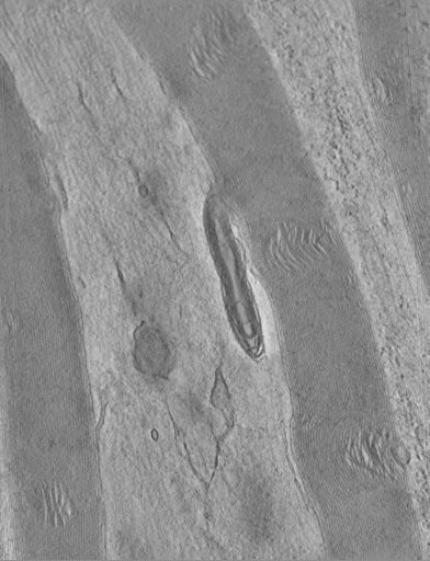
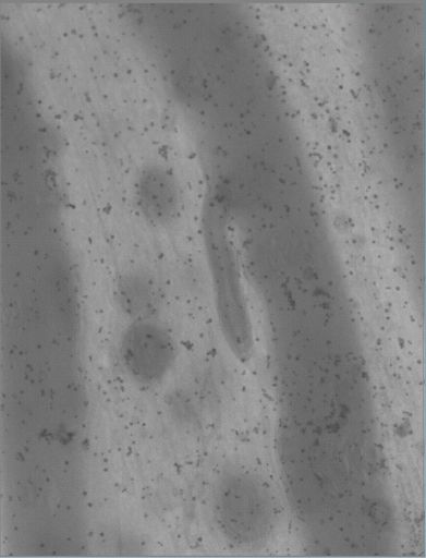
Single tilt image from a single axis tilt series through mitochondria at the Node of Ranvier of peripheral nerve root from rat. The dark speckles are gold particles that were applied to the surfaces to serve as fiducial marks for subsequent alignment.
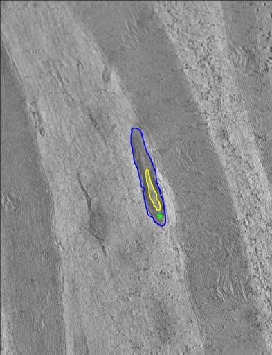
Manual segmentation of mitochondrial outer membrane and cristae using Xvoxtrace 2.17
Segmentation file description
Zip file containing the manual tracings in Xvoxtrace (.trace) along with the surface rendered objects in synu format (.synu) and the Viewdata file required by Synu. Segementations were performed on the Analyze 7.5 version of the reconstruction available from the reconstruction download section. The Xvoxtrace and Synu programs that were used to create this data are no longer supported. The Jinx tool (see CCDB tool list) can read the Xvoxtrace format and export the surfaces in multiple formats.
Study of morphology of quantitative morphology of mitochondria in the peripheral nerve of the adult rat using 3D electron microscopy
Funding agency
National Institute of Neurological Disorders and Stroke, National Center for Research Resources, National Institute of Diabetes and Kidney Diseases, National Library of Medicine
Leader(s)
Mark H. Ellisman
Guy A. Perkins
Start date
unspecified
End date
05-18-2010
Experiment
Experiment ID
8114
Title
Experiment 1
Purpose
Comparison of high pressure freezing and conventional fixation on mitochondrial structure using electron tomography
Experimenter(s)
Guy Perkins
Microscopy product
Microscopy product ID
8139
Instrument
JEOL4000EX
Microscopy type
IVEM
Product type
SINGLE TILT
Image basename
hpf3
Subject
Species
Rat
Scientific name
Rattus norvegicus
Strain
Sprague Dawley
Age
1 months
Age class
young adult
Tissue section
Anatomical location
spinal root
Microtome
Ultramicrotome
Specimen description
Organ
spinal root
System
peripheral nervous system
Structure
mitochondrion
Imaging parameters
Type
Electron microscopy product
Magification
0
Specimen preparation
Protocol used
Chemical fixation with 2% paraformaldehyde and 2.5% glutaraldehyde via vascular perfusion in 0.15M cacodylate buffer (pH 7.4) at 37 degrees C followed by high pressure freezing and freeze substitution using a Bal Tec HPM010 high pressure freezer (from Perkins et al., J. Structural Biology 161, 469, 2008).
Imaging product type
Type
Single tilt
Description
singlet_desc
Min range
-60 degrees
Max range
60 degrees
Tilt increment
2 degrees
Citation Information
Mark H. Ellisman, Guy A. Perkins CCDB:8139, Rattus norvegicus, mitochondrion. CIL. Dataset. https://doi.org/doi:10.7295/W9CCDB8139