Alternate header for print version
Advanced search
Contributors
Help
Submit
Search
menu
Cell Process
Cell Component
Cell Type
Organism
Microbial
Alzheimer's
Data Sets
Center for Research in Biological Systems
University of California, San Diego
9500 Gilman Drive
La Jolla, CA 92093-0608, USA
Voice
: (858) 534-0276
Fax
: (858) 534-7497
Email
: dorloff@ncmir.ucsd.edu
Search Results for
mesenchymal cell
(1945 results)
(Not the results you were expecting?)
(Comments)
CIL:39787
NCBI Organism Classification
Lytechinus pictus
Biological Process
embryonic morphogenesis
Cellular Component
filopodium
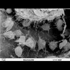
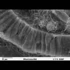
Scanning electron microscope image of Strongylocentrotus drobachiensus [sea urchin] at the gastrula stage. Embryo was split open to reveal the blastocoel cavity. There are several migrating mesenchyme...
CIL:39785
NCBI Organism Classification
Lytechinus pictus
Biological Process
embryonic morphogenesis
Cellular Component
cilium
Scanning electron microscope image of Strongylocentrotus drobachiensus [sea urchin] embryo at the late gastrula stage. Embryo was cut to reveal blastocoel cavity. The fertilization envelope dissolves ...
CIL:13091
NCBI Organism Classification
Mus musculus
Biological Process
immune system process
Cellular Component
microvillus
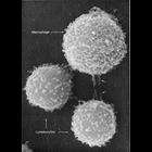
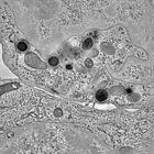
This image shows two different types of white blood cells from a mouse which are essential for mammalian immune response to protect from infection. The larger cell is a macrophage and the smaller two...
CIL:11634
NCBI Organism Classification
Rattus sp.
Biological Process
stereocilium
Cellular Component
cilium
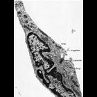
Figure 322 from Chapter 13 (Cilia and Flagella) of 'The Cell' by Don W. Fawcett M.D. Cilia and flagella usually occur on motile cells or on the free surfaces of epithelia. However, a solitary cilium i...
CIL:39786
NCBI Organism Classification
Lytechinus pictus
Biological Process
embryonic morphogenesis
Cellular Component
cilium
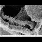
Scanning electron microscope image of Strongylocentrotus drobachiensus [sea urchin] at the gastrula stage. Embryo was split open to reveal a nice cross-section through the outer epithelial layer and t...
CIL:40011
NCBI Organism Classification
Homo sapiens
Biological Process
none specified
Cellular Component
immunological synapse
Tomogram of the immunological synapse between a cytotoxic T lymphocyte (CTL, top) and a target cell (bottom). The microtubule organization centre (MTOC) is polarized to the cell-cell contact site. The...
CIL:50301
NCBI Organism Classification
Mus musculus
Biological Process
none specified
Cellular Component
nucleus
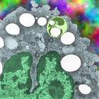
Transmission electron micrograph of an alveolar macrophage which contains E2, a fluorocarbon. This macrophage is from a mouse which liquid breathed that fluorocarbon for 3 hours and was allowed to rec...
CIL:38973
NCBI Organism Classification
none specified
Biological Process
histamine secretion
Cellular Component
histamine granules
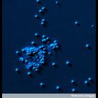
Confocal micrograph of a stimulated mast cell that has just exploded releasing numerous histamine granules. The remains of the cell are seen towards the lower left, still containing a number of granul...
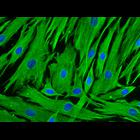
CIL:48101
NCBI Organism Classification
Homo sapiens
Biological Process
none specified
Cellular Component
Intermediate filament,
Cryopreserved human hair follicle dermal papilla cells were revived and stained for vimentin (in green) to reveal cytoskeleton, and DAPI (in blue) to label nuclei.
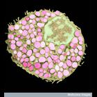
CIL:38944
NCBI Organism Classification
none specified
Biological Process
histamine transport
Cellular Component
histamine granules
Colorized transmission electron microscope image of a mast cell full of histamine granules (pink). Histamine is released as part of an allergic reaction. The diameter of the cell is approx. 8.4 microm...
« Previous
1
...
3
4
5
6
7
8
9
10
...
195
Next »
Results per page:
10
20
50
100