Alternate header for print version
Advanced search
Contributors
Help
Submit
Search
menu
Cell Process
Cell Component
Cell Type
Organism
Microbial
Alzheimer's
Data Sets
Center for Research in Biological Systems
University of California, San Diego
9500 Gilman Drive
La Jolla, CA 92093-0608, USA
Voice
: (858) 534-0276
Fax
: (858) 534-7497
Email
: dorloff@ncmir.ucsd.edu
Search Results for
neuron
(876 results)
(Not the results you were expecting?)
(Comments)
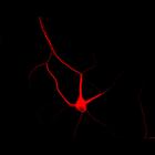
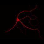
CIL:10311
NCBI Organism Classification
Rattus
Biological Process
cellular developmental process
Cellular Component
dendrite
Cultured hippocampal neurons after 10 days in vitro, immunostained for MAP2, a microtubule associated protein localized to dendrites (red) but not axons, which are not apparent in the immunofluorescen...
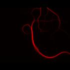
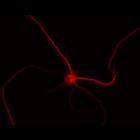
CIL:10318
NCBI Organism Classification
Rattus
Biological Process
cellular developmental process
Cellular Component
dendrite
Cultured hippocampal neurons after 10 days in vitro, immunostained for MAP2, a microtubule associated protein localized to dendrites (red) but not axons, which are not apparent in the immunofluorescen...
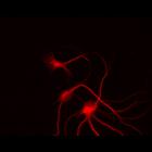
CIL:10324
NCBI Organism Classification
Rattus
Biological Process
cellular developmental process
Cellular Component
dendrite
Cultured hippocampal neurons after 10 days in vitro, immunostained for MAP2, a microtubule associated protein localized to dendrites (red) but not axons, which are not apparent in the immunofluorescen...
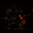
CIL:10114
NCBI Organism Classification
Rattus
Biological Process
developmental process
Cellular Component
cytoskeleton
This multi-layer image shows the spatial relationship between filamentous actin (red) and microtubule array (green) in cultured hippocampal neurons, grown for 5 days in vitro. Actin staining (with rh...
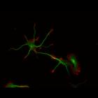
CIL:10116
NCBI Organism Classification
Rattus
Biological Process
developmental process
Cellular Component
cytoskeleton
This multi-layer image shows the spatial relationship between filamentous actin (red) and microtubule array (green) in cultured hippocampal neurons, grown for 5 days in vitro. Actin staining (with rh...
CIL:10119
NCBI Organism Classification
Rattus
Biological Process
developmental process
Cellular Component
cytoskeleton
This multi-layer image shows the spatial relationship between filamentous actin (red) and microtubule array (green) in cultured hippocampal neurons, grown for 5 days in vitro. Actin staining (with rh...
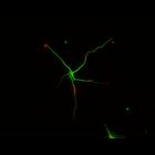
CIL:10210
NCBI Organism Classification
Rattus
Biological Process
developmental process
Cellular Component
cytoskeleton
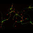
This multi-layer image shows the spatial relationship between filamentous actin (red) and microtubule array (green) in cultured hippocampal neurons, grown for 1 day in vitro. Actin staining (with rho...
CIL:10216
NCBI Organism Classification
Rattus
Biological Process
developmental process
Cellular Component
cytoskeleton
This multi-layer image shows the spatial relationship between filamentous actin (red) and microtubule array (green) in cultured hippocampal neurons, grown for 1 day in vitro. Actin staining (with rho...
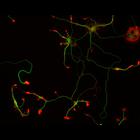
CIL:10355
NCBI Organism Classification
Rattus
Biological Process
cellular developmental process
Cellular Component
dendrite
Cultured hippocampal neurons after 14 days in vitro, immunostained for MAP2, a microtubule associated protein localized to dendrites (red) but not axons, which are not apparent in the immunofluorescen...
CIL:10357
NCBI Organism Classification
Rattus
Biological Process
cellular developmental process
Cellular Component
dendrite
Cultured hippocampal neurons after 14 days in vitro, immunostained for MAP2, a microtubule associated protein localized to dendrites (red) but not axons, which are not apparent in the immunofluorescen...
« Previous
1
...
81
82
83
84
85
86
87
88
Next »
Results per page:
10
20
50
100