Alternate header for print version
Advanced search
Contributors
Help
Submit
Search
menu
Cell Process
Cell Component
Cell Type
Organism
Microbial
Alzheimer's
Data Sets
Center for Research in Biological Systems
University of California, San Diego
9500 Gilman Drive
La Jolla, CA 92093-0608, USA
Voice
: (858) 534-0276
Fax
: (858) 534-7497
Email
: dorloff@ncmir.ucsd.edu
Search Results for
differential interference contrast ...
(1012 results)
(Not the results you were expecting?)
(Comments)
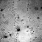
CIL:13871
NCBI Organism Classification
Saccharomyces cerevisiae
Biological Process
regulation of exit from mitosis
Cellular Component
nucleus
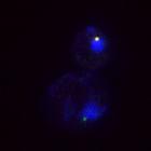
Tem1 (green) primarily localizes to the daughter cell spindle pole body (SPB) during anaphase. Control image for CIL# 13870 in which Tem1 loading onto SPBs was severely affected in bfa1Δ cells. DAPI ...
CIL:13885
NCBI Organism Classification
Saccharomyces cerevisiae
Biological Process
regulation of exit from mitosis
Cellular Component
nucleus
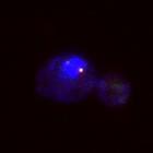
Tem1 normally localizes preferentially to the spindle pole body (SPB) that enters the daughter cell during anaphase (this image and CIL# 13882). This image shows SPB localization of eGFP-Tem1 (eGFP, g...
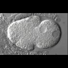
CIL:35126
NCBI Organism Classification
Caenorhabditis elegans
Biological Process
mitosis
Cellular Component
nucleus
This time-lapse series illustrates the early division up to the four-cell stage of a C. elegans embryo depleted of ufd-1 through RNAi. The first C. elegans embryonic division of the P0 zygote is asymm...
CIL:44506
NCBI Organism Classification
Canis lupus familiaris
Biological Process
wound healing, spreading of epidermal cells
Cellular Component
none specified
Time series light microscopy images illustrating a wound healing assay. A monolayer of MDCK (Madin-Darby Canine Kidney) epithelial cells is scratched to create a 'wound' about 300 micrometers in width...
CIL:44510
NCBI Organism Classification
Canis lupus familiaris
Biological Process
wound healing, spreading of epidermal cells
Cellular Component
none specified
Time series light microscopy images illustrating a wound healing assay. A monolayer of MDCK (Madin-Darby Canine Kidney) epithelial cells is scratched to create a 'wound' about 300 micrometers in width...
CIL:25630
NCBI Organism Classification
Caenorhabditis elegans
Biological Process
mitosis
Cellular Component
nucleus
This movie illustrates the early division up to the four-cell stage of a C. elegans embryo depleted of cdc-48 through RNAi. The first C. elegans embryonic division of the P0 zygote is asymmetric and g...
CIL:25631
NCBI Organism Classification
Caenorhabditis elegans
Biological Process
mitosis
Cellular Component
nucleus
This movie illustrates the early division up to the four-cell stage of a C. elegans embryo depleted of ufd-1 through RNAi. The first C. elegans embryonic division of the P0 zygote is asymmetric and ge...
CIL:25633
NCBI Organism Classification
Caenorhabditis elegans
Biological Process
mitosis
Cellular Component
nucleus
This movie illustrates the early division up to the four-cell stage of a C. elegans embryo depleted of cdc-48 and atl-1 through RNAi. The first C. elegans embryonic division of the P0 zygote is asymme...
CIL:13868
NCBI Organism Classification
Saccharomyces cerevisiae
Biological Process
cell cycle
Cellular Component
nucleus
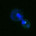
During late anaphase, Tem1 (green) localized to both spindle pole bodies (SPBs), but Bfa1 (red) was asymmetrically localized to the daughter SPB. DAPI (blue) and DIC (hidden) are also shown. Compare t...
CIL:13869
NCBI Organism Classification
Saccharomyces cerevisiae
Biological Process
cell cycle
Cellular Component
nucleus
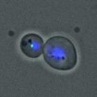
Both Tem1 (green) and Bfa1 (red) are localized to both spindle pole bodies in metaphase. DAPI (blue) and DIC (hidden) are also shown. Compare to anaphase image CIL# 13868. Image is Fig 6A, top panels,...
« Previous
1
...
94
95
96
97
98
99
100
101
102
Next »
Results per page:
10
20
50
100


")