Alternate header for print version
Advanced search
Contributors
Help
Submit
Search
menu
Cell Process
Cell Component
Cell Type
Organism
Microbial
Alzheimer's
Data Sets
Center for Research in Biological Systems
University of California, San Diego
9500 Gilman Drive
La Jolla, CA 92093-0608, USA
Voice
: (858) 534-0276
Fax
: (858) 534-7497
Email
: dorloff@ncmir.ucsd.edu
Search Results for
Cavia sp
(243 results)
(Not the results you were expecting?)
(Comments)
CIL:9903
NCBI Organism Classification
Euplotes sp.
Biological Process
cortical cytoskeleton organization
Cellular Component
microtubule basal body
Image of a dikinetid of Euplotes shows the transverse microtubular ribbon as well as a postciliary ribbon extending from the posterior basal body. Vesicles containing electron opaque bodies are typica...
CIL:9907
NCBI Organism Classification
Euplotes sp.
Biological Process
water transport
Cellular Component
contractile vacuole pore
A rare micrograph of the contractile vacuole and intact contractile vacuole pore membrane of Euplotes. Close examination at high magnification reveals the organization of microtubules lining the pore ...
CIL:41654
NCBI Organism Classification
Sedum sp
Biological Process
epidermis morphogenesis
Cellular Component
none specified
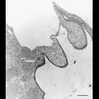
Brightfield image of the epidermis of Sedum sp. (stonecrop), a genus of flowering plants with succulent leaves. The image was taken at 125X. Honorable Mention, 2011 Olympus BioScapes Digital Imaging...
CIL:10773
NCBI Organism Classification
Cavia porcellus
Biological Process
translation
Cellular Component
endoplasmic reticulum
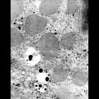
Figure 175 from Chapter 5 (Endoplasmic Reticulum) of 'The Cell, 2nd Ed.' by Don W. Fawcett M.D. Electron micrograph of a plasma cell from guinea pig bone marrow. Active synthesis of immunoglobulin b...
CIL:35998
NCBI Organism Classification
Homo sapiens
Biological Process
secretory granule organization
Cellular Component
secretory granule
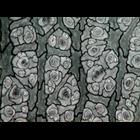
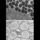
Figures 377 (upper) and 378 (lower) from Chapter 15 (Cytoplasmic Inclusions) of 'The Cell, 2nd Ed.' by Don W. Fawcett M.D. Comparison between the human pancreatic acinar cell (upper) and guinea pig pa...
CIL:37145
NCBI Organism Classification
Cavia porcellus
Biological Process
exocytosis
Cellular Component
exocytic vesicle
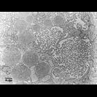
Transmission electron micrograph of exocytosis in a guinea pig pancreas. Image made available by James D. Jamieson and the Department of Cell Biology, Yale University School of Medicine.
CIL:37146
NCBI Organism Classification
Cavia porcellus
Biological Process
exocytosis
Cellular Component
exocytic vesicle
Transmission electron micrograph of exocytosis in a guinea pig pancreas. Image made available by James D. Jamieson and the Department of Cell Biology, Yale University School of Medicine.
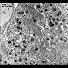
CIL:37230
NCBI Organism Classification
Cavia porcellus
Biological Process
organelle organization
Cellular Component
rough endoplasmic reticulum
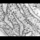
Classic early transmission electron micrograph of a thin section of guinea pig pancreas showing ribosome-studded rough endoplasmic reticulum. Image made available by James D. Jamieson and the Depart...
CIL:37232
NCBI Organism Classification
Cavia porcellus
Biological Process
organelle organization
Cellular Component
intracisternal granule
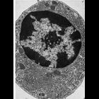
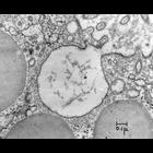
Early transmission electron micrograph showing intracisternal granules with an electron density equal to that of zymogen granules found in rough endoplasmic the reticulum lumen (cisternae) of animals ...
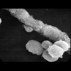
CIL:37317
NCBI Organism Classification
Cavia porcellus
Biological Process
none specified
Cellular Component
cell surface
Scanning electron micrograph of single guinea pic pancreatic acinar cells attached to a duct. Image made available by James D. Jamieson and the Department of Cell Biology, Yale University School of ...
« Previous
1
2
3
4
5
6
7
8
9
...
25
Next »
Results per page:
10
20
50
100