Alternate header for print version
Advanced search
Contributors
Help
Submit
Search
menu
Cell Process
Cell Component
Cell Type
Organism
Microbial
Alzheimer's
Data Sets
Center for Research in Biological Systems
University of California, San Diego
9500 Gilman Drive
La Jolla, CA 92093-0608, USA
Voice
: (858) 534-0276
Fax
: (858) 534-7497
Email
: dorloff@ncmir.ucsd.edu
Search Results for
differences in deposition of metal ...
(687 results)
(Not the results you were expecting?)
(Comments)
CIL:38811
NCBI Organism Classification
none specified
Biological Process
none specified
Cellular Component
cell surface
Scanning electron micrograph of red blood cells clearly showing their biconcave disc shape. Human red blood cells are typically 8 microns x 2 microns in size.
CIL:39104
NCBI Organism Classification
none specified
Biological Process
cell-cell adhesion
Cellular Component
cell surface
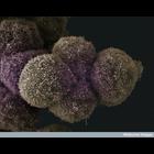
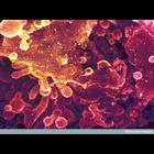
Scanning electron micrograph of pancreatic cancer cells. See additional image at CIL:39076.
CIL:39106
NCBI Organism Classification
none specified
Biological Process
none specified
Cellular Component
cell surface
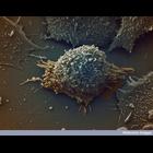
Colorized scanning electron micrograph of a lung cancer cell.
CIL:39358
NCBI Organism Classification
Zea mays
Biological Process
none specified
Cellular Component
none specified
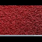
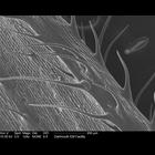
Scanning electron microscope image of corn leaf surface.
CIL:38945
NCBI Organism Classification
none specified
Biological Process
carbohydrate biosynthetic process
Cellular Component
Golgi stack
Colorized scanning electron micrograph showing the stacked membrane discs of the Golgi complex. The Golgi is the area within a cell where many carbohydrates are synthesised, which can be used to modif...
CIL:40652
NCBI Organism Classification
none specified
Biological Process
response to bacterium
Cellular Component
cell surface
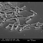
Scanning electron micrograph illustrating bacteria contamination of cells.
CIL:40656
NCBI Organism Classification
Bacillariophyta
Biological Process
cell wall organization
Cellular Component
frustule
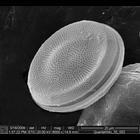
Scanning electron micrograph of a diatom showing the two silica based frustule (cell walls). Image also illustrates the regular pattern on the frustule.
CIL:40607
NCBI Organism Classification
Myxogastria
Biological Process
none specified
Cellular Component
cell surface
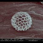
Scanning electron micrograph of a myxomycetes (a type of slime mold). This image was collected as part of a study to assess the diversity of myxomycetes in the Atlantic Forest.
CIL:41473
NCBI Organism Classification
coral
Biological Process
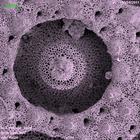
skeleton organization
Cellular Component
calcium skeleton
Scanning electron micrograph of the calcium skeleton of coral polyp. The sample was cleaned with bleach solution, dried and carbon coated prior to imaging. The image was collected using a secondary...
CIL:10427
NCBI Organism Classification
Gallus gallus
Biological Process
none specified
Cellular Component
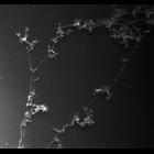
nuclear chromatin
Nucleated erythrocytes from chicken were spread on water, the dispersed chromatin picked up on carbon-formvar grids, fixed with paraformaldehye, critical point dried and shadowed with platinum. Image...
« Previous
1
2
3
4
5
6
7
8
9
...
69
Next »
Results per page:
10
20
50
100