Alternate header for print version
Advanced search
Contributors
Help
Submit
Search
menu
Cell Process
Cell Component
Cell Type
Organism
Microbial
Alzheimer's
Data Sets
Center for Research in Biological Systems
University of California, San Diego
9500 Gilman Drive
La Jolla, CA 92093-0608, USA
Voice
: (858) 534-0276
Fax
: (858) 534-7497
Email
: dorloff@ncmir.ucsd.edu
Search Results for
(15720 results)
(Not the results you were expecting?)
(Comments)
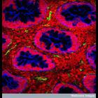
CIL:38952
NCBI Organism Classification
Homo sapiens
Biological Process
Gram-negative bacterial cell surface binding
Cellular Component
Shiga toxin
A confocal micrograph of an intestinal biopsy from a child infected with shiga toxin-producing E. coli. Shiga toxin is an extremely potent toxin that is produced when the bacterium contains a bacteri...
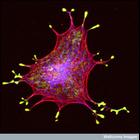
CIL:38956
NCBI Organism Classification
none specified
Biological Process
melanosome localization
Cellular Component
melanosome
Confocal micrograph of an isolated melanin-producing cell (a melanocyte) showing the melanosomes (vesicles that hold the melanin granules) in yellow, the actin in red and the microtubules in blue. The...
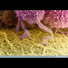
CIL:38957
NCBI Organism Classification
Homo sapiens
Biological Process
receiving nourishment
Cellular Component
zona pellucida
A colorized scanning electron micrograph of a human egg, which is the huge cell colored yellow at the bottom of this image. The follicle cells that surround it (top) send out long projections that pen...
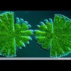
CIL:38959
NCBI Organism Classification
Micrasterias rotata
Biological Process
cytokinesis by binary fission
Cellular Component
cell surface
Differential interference micrograph (Nomarski) of late binary fission in the desmid algae, Micrasterias rotata.
CIL:38963
NCBI Organism Classification
Drosophila
Biological Process
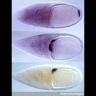
intracellular mRNA localization
Cellular Component
oskar mRNA
In situ hybridizations showing the localization of bicoid (top), oskar (middle) and gurken (bottom) mRNAs in the stage 10 Drosophila egg chambers. These localizations define the anterior- posterior a...
CIL:38978
NCBI Organism Classification
Homo sapiens
Biological Process
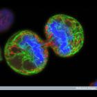
mitosis
Cellular Component
endoplasmic reticulum
Human melanoma cell undergoing cell division. The chromosomes (blue) have separated and the two daughter cells have almost split apart - only a small bridge of cytoplasm remains. The green staining la...
CIL:38980
NCBI Organism Classification
Gallus gallus
Biological Process
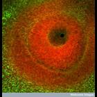
eye development
Cellular Component
cell surface
Confocal image of the developing eye of a chick embryo. The dark region in the centre is the where the surface layer has invaginated to form the lens vesicle. The tissue surrounding this space is stai...
CIL:38995
NCBI Organism Classification
Homo sapiens
Biological Process
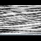
collagen fibril organization
Cellular Component
extracellular matrix
Transmission electron micrograph showing collagen fibrils in longitudinal view from a patient with glaucoma. The a-e banding is visualized by uranyl acetate staining of charged amino acid residues in ...
CIL:39003
NCBI Organism Classification
Mus musculus
Biological Process
mitosis
Cellular Component
actin cytoskeleton
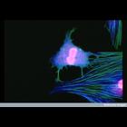
Confocal microscope image of a mouse fibroblast cell about to divide. The nuclei are stained red, and the actin and tubulin filaments of the cytoskeleton, forming the internal framework, are stained g...
CIL:39006
NCBI Organism Classification
Homo sapiens
Biological Process
blastocyst hatching
Cellular Component
zona pellucida
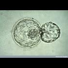
Light micrograph of a human embryo at the blastocyst stage, about six days after fertilization. The embryo is in the process of "hatching" out of the zona pellucida - the tough outer membrane - just b...
« Previous
1
...
1072
1073
1074
1075
1076
1077
1078
1079
...
1572
Next »
Results per page:
10
20
50
100