Alternate header for print version
Advanced search
Contributors
Help
Submit
Search
menu
Cell Process
Cell Component
Cell Type
Organism
Microbial
Alzheimer's
Data Sets
Center for Research in Biological Systems
University of California, San Diego
9500 Gilman Drive
La Jolla, CA 92093-0608, USA
Voice
: (858) 534-0276
Fax
: (858) 534-7497
Email
: dorloff@ncmir.ucsd.edu
Search Results for
microtubule plus end
(3382 results)
(Not the results you were expecting?)
(Comments)
CIL:6158
NCBI Organism Classification
Rattus
Biological Process
neuron development
Cellular Component
cytoskeleton
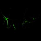
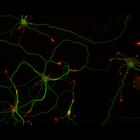
Synapse formation in cultured hippocampal neurons after 7 days in vitro. Cells were immunostained for MAP2, a microtubule associated protein localized to dendrites but not axons (green), and synapsin...
CIL:6166
NCBI Organism Classification
Rattus
Biological Process
neuron development
Cellular Component
cytoskeleton
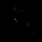
Synapse formation in cultured hippocampal neurons after 7 days in vitro. Cells were immunostained for MAP2, a microtubule associated protein localized to dendrites but not axons (green), and synapsin...
CIL:6203
NCBI Organism Classification
Rattus
Biological Process
neuron development
Cellular Component
cytoskeleton
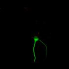
Synapse formation in cultured hippocampal neurons after 7 days in vitro. Cells were immunostained for MAP2, a microtubule associated protein localized to dendrites but not axons (green), and synapsin...
CIL:6215
NCBI Organism Classification
Rattus
Biological Process
neuron development
Cellular Component
cytoskeleton
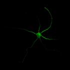
Synapse formation in cultured hippocampal neurons after 7 days in vitro. Cells were immunostained for MAP2, a microtubule associated protein localized to dendrites but not axons (green), and synapsin...
CIL:10113
NCBI Organism Classification
Rattus
Biological Process
developmental process
Cellular Component
cytoskeleton
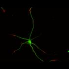
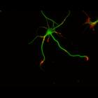
This multi-layer image shows the spatial relationship between filamentous actin (red) and microtubule array (green) in cultured hippocampal neurons, grown for 5 days in vitro. Actin staining (with rh...
CIL:10208
NCBI Organism Classification
Rattus
Biological Process
developmental process
Cellular Component
cytoskeleton
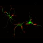
This multi-layer image shows the spatial relationship between filamentous actin (red) and microtubule array (green) in cultured hippocampal neurons, grown for 1 day in vitro. Actin staining (with rho...
CIL:10211
NCBI Organism Classification
Rattus
Biological Process
developmental process
Cellular Component
cytoskeleton
This multi-layer image shows the spatial relationship between filamentous actin (red) and microtubule array (green) in cultured hippocampal neurons, grown for 1 day in vitro. Actin staining (with rho...
CIL:10218
NCBI Organism Classification
Rattus
Biological Process
developmental process
Cellular Component
cytoskeleton
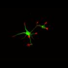
This multi-layer image shows the spatial relationship between filamentous actin (red) and microtubule array (green) in cultured hippocampal neurons, grown for 3 days in vitro. Actin staining (with rh...
CIL:10224
NCBI Organism Classification
Rattus
Biological Process
developmental process
Cellular Component
cytoskeleton
This multi-layer image shows the spatial relationship between filamentous actin (red) and microtubule array (green) in cultured hippocampal neurons, grown for 3 days in vitro. Actin staining (with rh...
CIL:12289
NCBI Organism Classification
Homo sapiens
Biological Process
cell-substrate adhesion
Cellular Component
microtubule
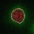
To determine the extent to which extracellular matrix proteins colocalize with the microtubule-anchoring factor LL5β, MCF-10A epithelial cells were immunostained with antibodies to the secreted autoc...
« Previous
1
...
9
10
11
12
13
14
15
16
...
339
Next »
Results per page:
10
20
50
100