Alternate header for print version
Advanced search
Contributors
Help
Submit
Search
menu
Cell Process
Cell Component
Cell Type
Organism
Microbial
Alzheimer's
Data Sets
Center for Research in Biological Systems
University of California, San Diego
9500 Gilman Drive
La Jolla, CA 92093-0608, USA
Voice
: (858) 534-0276
Fax
: (858) 534-7497
Email
: dorloff@ncmir.ucsd.edu
Search Results for
muscle contraction
(265 results)
(Not the results you were expecting?)
(Comments)
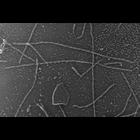
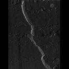
CIL:6265
NCBI Organism Classification
Oryctolagus cuniculus
Biological Process
skeletal muscle contraction
Cellular Component
cytoskeleton
Rabbit psoas skeletal muscle fibers were blended in the presence of 5 mM MgATP to dissociate thick and thin filaments. The medium was then gently replaced with 0.3M KCl to dissociate thick filaments....
CIL:6268
NCBI Organism Classification
Oryctolagus cuniculus
Biological Process
skeletal muscle contraction
Cellular Component
cytoskeleton
Rabbit psoas skeletal muscle fibers were blended in the presence of 5 mM MgATP to dissociate thick and thin filaments. The medium was then gently replaced with 0.3M KCl to dissociate thick filaments....
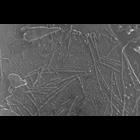
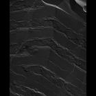
CIL:832
NCBI Organism Classification
Oryctolagus cuniculus
Biological Process
skeletal muscle contraction
Cellular Component
cytoskeleton
Purified rabbit skeletal muscle myosin in 0.6M KCl was deposited onto mica flakes, quick-frozen by contact with a liquid helium- cooled copper block in a Heuser-type cryopress, and freeze-etched in a ...
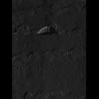
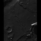
CIL:18442
NCBI Organism Classification
damselfly
Biological Process
skeletal muscle contraction
Cellular Component
myofibril
Most images show fractures through the outer membrane of mitochondria and SR/T tubules of dyads nestled in long grooves of the mitochondrial surface. All leaflets of the membranes appear in the image...
CIL:18446
NCBI Organism Classification
damselfly
Biological Process
skeletal muscle contraction
Cellular Component
myofibril
Most images show fractures through the outer membrane of mitochondria and SR/T tubules of dyads nestled in long grooves of the mitochondrial surface. All leaflets of the membranes appear in the image...
CIL:18483
NCBI Organism Classification
damselfly
Biological Process
skeletal muscle contraction
Cellular Component
myofibril
Most images show fractures through the outer membrane of mitochondria and SR/T tubules of dyads nestled in long grooves of the mitochondrial surface. All leaflets of the membranes appear in the image...
CIL:18459
NCBI Organism Classification
damselfly
Biological Process
skeletal muscle contraction
Cellular Component
myofibril
Most images show fractures through the outer membrane of mitochondria and SR/T tubules of dyads nestled in long grooves of the mitochondrial surface. All leaflets of the membranes appear in the image...
CIL:18465
NCBI Organism Classification
damselfly
Biological Process
skeletal muscle contraction
Cellular Component
myofibril
Most images show fractures through the outer membrane of mitochondria and SR/T tubules of dyads nestled in long grooves of the mitochondrial surface. All leaflets of the membranes appear in the image...
CIL:18471
NCBI Organism Classification
damselfly
Biological Process
skeletal muscle contraction
Cellular Component
myofibril
Most images show fractures through the outer membrane of mitochondria and SR/T tubules of dyads nestled in long grooves of the mitochondrial surface. All leaflets of the membranes appear in the image...
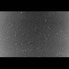
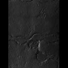
CIL:2873
NCBI Organism Classification
Oryctolagus cuniculus
Biological Process
skeletal muscle contraction
Cellular Component
cytoskeleton
Rabbit psoas skeletal muscle fibers were held at constant length and rigor induced by immersion in 0.2% saponin. The fibers were quick-frozen by contact with a liquid helium-cooled copper block in a ...
« Previous
1
...
14
15
16
17
18
19
20
21
...
27
Next »
Results per page:
10
20
50
100