Alternate header for print version
Advanced search
Contributors
Help
Submit
Search
menu
Cell Process
Cell Component
Cell Type
Organism
Microbial
Alzheimer's
Data Sets
Center for Research in Biological Systems
University of California, San Diego
9500 Gilman Drive
La Jolla, CA 92093-0608, USA
Voice
: (858) 534-0276
Fax
: (858) 534-7497
Email
: dorloff@ncmir.ucsd.edu
Search Results for
actin cytoskeleton
(655 results)
(Not the results you were expecting?)
(Comments)
CIL:34885
NCBI Organism Classification
Xenopus laevis
Biological Process
branching of actin filaments
Cellular Component
actin cytoskeleton
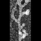
Multiple branching of actin filaments in lamellipodia of Xenopus keratocytes. This image show an enlargement of a local region from the overview of the leading edge, CIL 24786. Image corresponds to F...
CIL:34890
NCBI Organism Classification
none specified
Biological Process
branching of actin filaments
Cellular Component
actin cytoskeleton
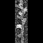
Multiple branching of actin filaments in lamellipodia of vertebrate fibroblasts. This image shows a local enlargement of the leading edge shown in overview in CIL 24788. Image corresponds to Figure 1...
CIL:35619
NCBI Organism Classification
Homo sapiens
Biological Process
cytoskeleton organization
Cellular Component
actin cytoskeleton
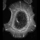
Actin fiber organization in primary keratinocyte. There are numerous actin arcs around the periphery of the cell and actin bundles that terminate in focal adhesions (bright structures perpendicular t...
CIL:35062
NCBI Organism Classification
none specified
Biological Process
branching of actin filaments
Cellular Component
actin cytoskeleton
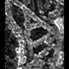
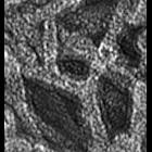
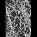
Electron micrograph of keratocyte or fibroblast lamellipodial actin network after unprotected extraction. All examples demonstrate frequent branching of actin filaments. Image corresponds to a singl...
CIL:35063
NCBI Organism Classification
none specified
Biological Process
branching of actin filaments
Cellular Component
actin cytoskeleton
Electron micrograph of keratocyte or fibroblast lamellipodial actin network after unprotected extraction. All examples demonstrate frequent branching of actin filaments. Image corresponds to a singl...
CIL:36070
NCBI Organism Classification
Mus musculus
Biological Process
actin cytoskeleton organization
Cellular Component
actin cytoskeleton
Figure 438 from Chapter 16 (Cytoplasmic matrix and cytoskeleton) of 'The Cell, 2nd Ed.' by Don W. Fawcett M.D. Two bundles of actin-rich stress fibers are apparent in this high magnification microgra...
CIL:36695
NCBI Organism Classification
Danio rerio
Biological Process
mitosis
Cellular Component
microtubule
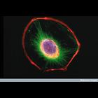
Confocal z-stack of a dividing zebrafish embryo. The fixed embryo is visualized using EMTB-3GFP (microtubules-marker - green), phalloidin (actin marker -red) and Hoechst (DNA marker - blue).
CIL:39004
NCBI Organism Classification
Mus musculus
Biological Process
cytoskeleton organization
Cellular Component
intermediate filament cytoskeleton
Confocal microscope image of a 3T3 fibroblast cell. The nucleus has been stained blue, and two components of the cytoskeleton, actin microfilaments and intermediate filaments, are stained red and gree...
CIL:40810
NCBI Organism Classification
Rattus
Biological Process
dendrite morphogenesis
Cellular Component
axon
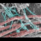
Colorized transmission electron micrograph of a platinum replica showing the cytoskeletal organization of stubby dendritic spines in extracted hippocampal neurons after 14 DIV. This imge shows branche...
CIL:42516
NCBI Organism Classification
Rattus
Biological Process
none specified
Cellular Component
actin cytoskeleton
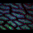
Fluorescence micrograph of the gut of transgenic GFP rat showing actin (red) and nuclei (blue). Image taken with a 20X objective. Honorable mention, 2004 Olympus BioScapes Digital Imaging Competitio...
« Previous
1
...
10
11
12
13
14
15
16
17
...
66
Next »
Results per page:
10
20
50
100


")