Alternate header for print version
Advanced search
Contributors
Help
Submit
Search
menu
Cell Process
Cell Component
Cell Type
Organism
Microbial
Alzheimer's
Data Sets
Center for Research in Biological Systems
University of California, San Diego
9500 Gilman Drive
La Jolla, CA 92093-0608, USA
Voice
: (858) 534-0276
Fax
: (858) 534-7497
Email
: dorloff@ncmir.ucsd.edu
Search Results for
actin cytoskeleton
(655 results)
(Not the results you were expecting?)
(Comments)
CIL:10215
NCBI Organism Classification
Rattus
Biological Process
developmental process
Cellular Component
cytoskeleton
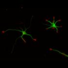
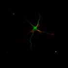
This multi-layer image shows the spatial relationship between filamentous actin (red) and microtubule array (green) in cultured hippocampal neurons, grown for 1 day in vitro. Actin staining (with rho...
CIL:10220
NCBI Organism Classification
Rattus
Biological Process
developmental process
Cellular Component
cytoskeleton
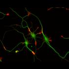
This multi-layer image shows the spatial relationship between filamentous actin (red) and microtubule array (green) in cultured hippocampal neurons, grown for 3 days in vitro. Actin staining (with rh...
CIL:10228
NCBI Organism Classification
Rattus
Biological Process
developmental process
Cellular Component
cytoskeleton
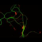
This multi-layer image shows the spatial relationship between filamentous actin (red) and microtubule array (green) in cultured hippocampal neurons, grown for 5 days in vitro. Actin staining (with rh...
CIL:8782
NCBI Organism Classification
Rattus
Biological Process
developmental process
Cellular Component
cytoskeleton
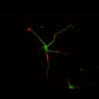
This color combined image shows the spatial relationship between filamentous actin (red) and microtubule array (green) in cultured hippocampal neurons, grown for 1 day in vitro. Actin staining (with ...
CIL:8775
NCBI Organism Classification
Rattus
Biological Process
developmental process
Cellular Component
cytoskeleton
This color combined image shows the spatial relationship between filamentous actin (red) and microtubule array (green) in cultured hippocampal neurons, grown for 1 day in vitro. Actin staining (with ...
CIL:12010
NCBI Organism Classification
Hypsophrys nicaraguensis
Biological Process
centripetal actin flow
Cellular Component
actin cytoskeleton
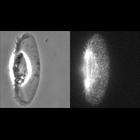
Visualization of F-actin network movement in stationary keratocytes with FSM. F-actin flows centripetally from the cell periphery to the cell body in stationary cells. Video shows paired phase-cont...
CIL:12011
NCBI Organism Classification
Hypsophrys nicaraguensis
Biological Process
actin flow
Cellular Component
actin cytoskeleton
Visualization of F-actin network movement in motile keratocytes with FSM. F-actin flows slowly from the leading edge to the cell body in motile cells. Video is paired phase-contrast and AF546-phallo...
CIL:12297
NCBI Organism Classification
Taricha granulosa
Biological Process
actin filament polymerization
Cellular Component
actin cytoskeleton
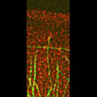
The retrograde flow of microtubules oriented perpendicular to the leading edge in the lamellipodium is coupled to the movement of immediately adjacent lamellum f-actin speckles. Primary cultures of n...
CIL:31923
NCBI Organism Classification
Saccharomyces cerevisiae
Biological Process
receptor-mediated endocytosis
Cellular Component
endocytic vesicle
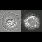
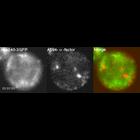
Localization of Alexa Fluor-594-α-factor-labeled endosomes (center; red in merge) and Abp140-3GFP (left; green in merge) in bni1-12 bnr1Δ cell. Abp140p binds F-actin and localizes to actin patches ...
CIL:37205
NCBI Organism Classification
Rattus
Biological Process
voluntary skeletal muscle contraction
Cellular Component
mitochondrion
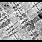
Transmission electron micrograph of rat sartorius striated muscle showing the regular arrangement of the actin-myosin myofilaments and abundant mitochondria. This 1956 TEM image has historical value...
« Previous
1
...
22
23
24
25
26
27
28
29
...
66
Next »
Results per page:
10
20
50
100