Alternate header for print version
Advanced search
Contributors
Help
Submit
Search
menu
Cell Process
Cell Component
Cell Type
Organism
Microbial
Alzheimer's
Data Sets
Center for Research in Biological Systems
University of California, San Diego
9500 Gilman Drive
La Jolla, CA 92093-0608, USA
Voice
: (858) 534-0276
Fax
: (858) 534-7497
Email
: dorloff@ncmir.ucsd.edu
Search Results for
cell surface
(1377 results)
(Not the results you were expecting?)
(Comments)
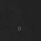
CIL:39649
NCBI Organism Classification
Saccharomyces cerevisiae
Biological Process
cell cycle checkpoint
Cellular Component
cell surface
10-hour timelapse of Wild-type Saccharomyces cerevisiae growing in 2% raffinose. These experiments are the control experiments used for comparison to the effects of the double mutant Clb2dbΔ, Clb5Δ ...
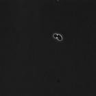
CIL:39651
NCBI Organism Classification
Saccharomyces cerevisiae
Biological Process
cell cycle checkpoint
Cellular Component
cell surface
10-hour timelapse of Wild-type Saccharomyces cerevisiae growing in 2% raffinose. These experiments are the control experiments used for comparison to the effects of the double mutant Clb2dbΔ, Clb5Δ ...
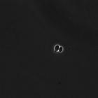
CIL:39656
NCBI Organism Classification
Saccharomyces cerevisiae
Biological Process
cell cycle checkpoint
Cellular Component
cell surface
10-hour timelapse of Wild-type Saccharomyces cerevisiae growing in 2% raffinose. These experiments are the control experiments used for comparison to the effects of the double mutant Clb2dbΔ, Clb5Δ ...
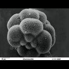
CIL:39783
NCBI Organism Classification
Lytechinus pictus
Biological Process
embryonic cleavage
Cellular Component
cell surface
Scanning electron microscope image of Lytechinus pictus [sea urchin] embryo at the 16-cell stage. The four large macromeres are behind the four small micromeres. The eight mesomeres are behind the mac...
CIL:39788
NCBI Organism Classification
Lytechinus pictus
Biological Process
embryonic morphogenesis
Cellular Component
cell surface
Scanning electron microscope image of Strongylocentrotus drobachiensus embryo at the primary mesenchyme blastula stage. Embryo was split open to reveal the outer epithelial layer and the blastocoel ca...
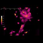
CIL:43451
NCBI Organism Classification
Homo sapiens
Biological Process
asymmetric cell division
Cellular Component
cell surface
Digital holographic microscopy video showing cell division of unlabeled JIMT-1 breast cancer cells. During the video, spanning over 72 hours, one cell divides into three daughter cells (seen on top of...
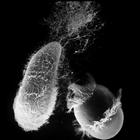
CIL:22781
NCBI Organism Classification
Didinium nasutum
Biological Process
response to toxin
Cellular Component
cell cortex
Didinium attacks Paramecium. Also showing many discharged trichocysts and metachronous waves of cilia in the two characteristic ciliary girdles of Didinium nasutum and on Paramecium. A series of captu...
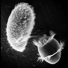
CIL:21993
NCBI Organism Classification
Didinium nasutum
Biological Process
phagocytosis
Cellular Component
oral apparatus
Didinium captures Paramecium. A moment after initial contact when toxicysts enter Paramecium. The strand of toxicysts can be seen between the two organisms, and Didinium will use the anchored toxicyst...
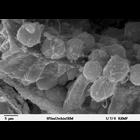
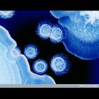
CIL:38807
NCBI Organism Classification
Capnocytophaga
Biological Process
none specified
Cellular Component
cell surface
This color-enhanced photomicrograph shows different species of bacteria that cause dental plaque - a colorless film that forms on teeth caused by the growth of bacterial colonies. Plaque develops natu...
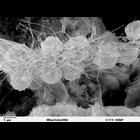
CIL:39789
NCBI Organism Classification
Lytechinus pictus
Biological Process
syncytial ring formation
Cellular Component
cell surface
Scanning electron microscope image of Strongylocentrotus drobachiensus [sea urchin] embryo at the late gastrula stage. This is a high magnification image of CIL 39785. Embryo was cut to reveal blastoc...
« Previous
1
...
17
18
19
20
21
22
23
24
...
138
Next »
Results per page:
10
20
50
100