Alternate header for print version
Advanced search
Contributors
Help
Submit
Search
menu
Cell Process
Cell Component
Cell Type
Organism
Microbial
Alzheimer's
Data Sets
Center for Research in Biological Systems
University of California, San Diego
9500 Gilman Drive
La Jolla, CA 92093-0608, USA
Voice
: (858) 534-0276
Fax
: (858) 534-7497
Email
: dorloff@ncmir.ucsd.edu
Search Results for
confocal microscopy
(2626 results)
(Not the results you were expecting?)
(Comments)
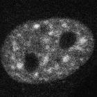
CIL:26594
NCBI Organism Classification
Homo sapiens
Biological Process
RNA splicing
Cellular Component
pre-mRNA
HeLa cells stably expressing BAC-encoded GFP-labeled snRNP protein hPrp8 and imaged witb a Leica SP5 confocal microscope using a 60x 1.4NA HCX Plan Apo objective lens. The hPrp8 is confined to the nu...
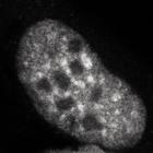
CIL:26597
NCBI Organism Classification
Homo sapiens
Biological Process
RNA splicing
Cellular Component
pre-mRNA
HeLa cells stably expressing the BAC-encoded GFP-labeled hPrp4 snRNP and MS2-mRED protein and treated with doxycholine to express the E3 transgene. Shown is the GFP signal. Cells imaged witb a Leica ...
CIL:26604
NCBI Organism Classification
Homo sapiens
Biological Process
RNA splicing
Cellular Component
pre-mRNA
HeLa cells stably expressing the BAC-encoded GFP-labeled U2B snRNP and MS2-mRED protein and treated with doxycholine to express the E3 transgene. Shown is the GFP signal. Cells imaged witb a Leica SP...
CIL:35161
NCBI Organism Classification
Homo sapiens
Biological Process
none specified
Cellular Component
mitotic membranes
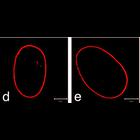
Live mitotic HeLa cell treated with control siRNA, DiOC6(3)to label mitotic membranes (green), and Hoechst 33258 to label chromosomes (blue). Confocal images were taken at 0.118 μm steps along the Z-...
CIL:36028
NCBI Organism Classification
Homo sapiens
Biological Process
none specified
Cellular Component
nuclear envelope
Shown are a normal human fibroblasts stained for lamin A (red), a major component of the nuclear lamina. Other images in this group show the envelope abnormalities that occur in HGPS patients carryin...
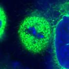
CIL:38956
NCBI Organism Classification
none specified
Biological Process
melanosome localization
Cellular Component
melanosome
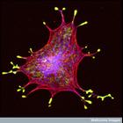
Confocal micrograph of an isolated melanin-producing cell (a melanocyte) showing the melanosomes (vesicles that hold the melanin granules) in yellow, the actin in red and the microtubules in blue. The...
CIL:38978
NCBI Organism Classification
Homo sapiens
Biological Process
mitosis
Cellular Component
endoplasmic reticulum
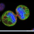
Human melanoma cell undergoing cell division. The chromosomes (blue) have separated and the two daughter cells have almost split apart - only a small bridge of cytoplasm remains. The green staining la...
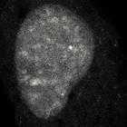
CIL:38980
NCBI Organism Classification
Gallus gallus
Biological Process
eye development
Cellular Component
cell surface
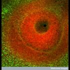
Confocal image of the developing eye of a chick embryo. The dark region in the centre is the where the surface layer has invaginated to form the lens vesicle. The tissue surrounding this space is stai...
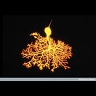
CIL:39003
NCBI Organism Classification
Mus musculus
Biological Process
mitosis
Cellular Component
actin cytoskeleton
Confocal microscope image of a mouse fibroblast cell about to divide. The nuclei are stained red, and the actin and tubulin filaments of the cytoskeleton, forming the internal framework, are stained g...
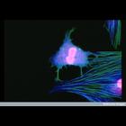
CIL:39010
NCBI Organism Classification
Rattus
Biological Process
cerebral Purkinje cell morphogenesis
Cellular Component
neuronal cell body
Confocal microscope image of a developing Purkinje cell in the cerebellum of a 9 day postnatal rat. The cerebellum contains millions of these cells which are involved with integrating motor and sensor...
« Previous
1
...
17
18
19
20
21
22
23
24
...
263
Next »
Results per page:
10
20
50
100

")