Alternate header for print version
Advanced search
Contributors
Help
Submit
Search
menu
Cell Process
Cell Component
Cell Type
Organism
Microbial
Alzheimer's
Data Sets
Center for Research in Biological Systems
University of California, San Diego
9500 Gilman Drive
La Jolla, CA 92093-0608, USA
Voice
: (858) 534-0276
Fax
: (858) 534-7497
Email
: dorloff@ncmir.ucsd.edu
Search Results for
confocal microscopy
(2626 results)
(Not the results you were expecting?)
(Comments)
CIL:25541
NCBI Organism Classification
Drosophila melanogaster
Biological Process
calcium-dependent cell-cell adhesion
Cellular Component
adherens junction
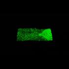
A time-lapse movie showing the dorsal pouch formation and migration of heart-anchoring cells (HANCs) in the Drosophila embryo expressing ubi-DE-cad-GFP (Shg-GFP). Shg-positive HANCs corresponding to t...
CIL:31203
NCBI Organism Classification
Gallus gallus
Biological Process
axonal transport
Cellular Component
mitochondrion
Mitochondrial transport during a growth cone pause. Chicken DRG neurons were grown on coverslips coated with laminin/poly-L-ornithine for 2 d and stained with 0.1 µM MitoTracker Red CMXRos. Images we...
CIL:13547
NCBI Organism Classification
Homo sapiens
Biological Process
Golgi organization
Cellular Component
Golgi stack
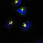
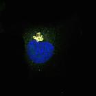
GalNac-T1 staining (red) and Helix Pomatia Lectin (HPL) (green) colocalizes exclusively at the Golgi (Giantin) (gray) in unstimulated HeLa cells. The Tn antigen refers to terminal α-linked N-acetyl g...
CIL:13557
NCBI Organism Classification
Homo sapiens
Biological Process
Golgi organization
Cellular Component
Golgi stack
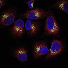
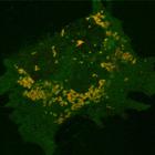
Helix Pomatia Lectin (HPL) (gray) staining is present exclusively at the Golgi (Giantin) (green) with no significant colocalization with ER marker, PDI (protein disulfide isomerase) (red) in unstimula...
CIL:13565
NCBI Organism Classification
Homo sapiens
Biological Process
retrograde vesicle-mediated transport, Golgi to ER
Cellular Component
Golgi cisterna membrane
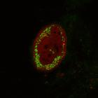
COP-I gamma1 (green) staining at the Golgi (GM130, a cis-Golgi marker) (red) in unstimulated HeLa cells. Helix Pomatia Lectin (HPL) (gray) staining is also shown. HPL binds various glycans but the Tn ...
CIL:13727
NCBI Organism Classification
Mus musculus
Biological Process
mitochondrion degradation
Cellular Component
mitochondrion
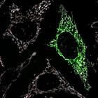
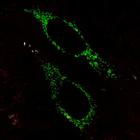
Upon mitochondrial uncoupling, wild-type PINK1-YFP (green) and mCherry-Parkin (red) are localized to mitochondria in PINK1 knock-out mouse embryonic fibroblasts (PINK1 KO MEFs). Transfected PINK1 KO M...
CIL:13728
NCBI Organism Classification
Mus musculus
Biological Process
mitochondrion degradation
Cellular Component
mitochondrion
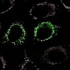
Upon mitochondrial uncoupling, transmembrane domain-deleted PINK1-YFP (green) fails to recruit mCherry-Parkin (red) to mitochondria in PINK1 knock-out mouse embryonic fibroblasts (PINK1 KO MEFs). Tran...
CIL:13729
NCBI Organism Classification
Homo sapiens
Biological Process
respiratory electron transport chain
Cellular Component
mitochondrion
Mitochondria in HeLa cells are identified by mito-YFP (green). When fixed cells are permeabilized with 0.005% digitonin, the antibody to cyctochrome c (red), a mitochondrial matrix marker, is unable t...
CIL:13730
NCBI Organism Classification
Homo sapiens
Biological Process
respiratory electron transport chain
Cellular Component
mitochondrion
Mitochondria in HeLa cells are identified by mito-YFP (green) following treatment with the mitochondrial depolarizing agent CCCP (carbonyl cyanide m-chlorophenyl hydrazone). When fixed cells are unper...
CIL:13731
NCBI Organism Classification
Homo sapiens
Biological Process
respiratory electron transport chain
Cellular Component
mitochondrion
Mitochondria in HeLa cells are identified by mito-YFP (green) following treatment with the mitochondrial depolarizing agent CCCP (carbonyl cyanide m-chlorophenyl hydrazone). When fixed cells are perme...
« Previous
1
...
254
255
256
257
258
259
260
261
...
263
Next »
Results per page:
10
20
50
100