Alternate header for print version
Advanced search
Contributors
Help
Submit
Search
menu
Cell Process
Cell Component
Cell Type
Organism
Microbial
Alzheimer's
Data Sets
Center for Research in Biological Systems
University of California, San Diego
9500 Gilman Drive
La Jolla, CA 92093-0608, USA
Voice
: (858) 534-0276
Fax
: (858) 534-7497
Email
: dorloff@ncmir.ucsd.edu
Search Results for
confocal microscopy
(2626 results)
(Not the results you were expecting?)
(Comments)
CIL:48808
NCBI Organism Classification
Mus musculus
Biological Process
Cancer
Cellular Component
none specified
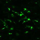
To determine the origin (local vs. circulating) of tumor associated fibroblasts, a parabiotic pair of mice was created where only one mouse expressed EGFP under the control of the type I collagen alph...
CIL:48810
NCBI Organism Classification
Mus musculus
Biological Process
Cancer
Cellular Component
none specified
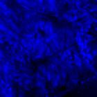
To determine the origin (local vs. circulating) of tumor associated fibroblasts, a parabiotic pair of mice was created where only one mouse expressed EGFP under the control of the type I collagen alph...
CIL:7669
NCBI Organism Classification
Mus musculus
Biological Process
none specified
Cellular Component
nuclear chromatin
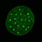
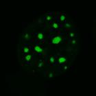
Fluorescence recovery after photobleaching (FRAP)kinetics of histone H1-zero were recorded using mouse fibroblast cell line stably expressing GFP-H10. Live cells were imaged using a Zeiss LSM 510 meta...
CIL:8047
NCBI Organism Classification
Mus musculus
Biological Process
none specified
Cellular Component
nuclear chromatin
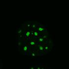
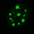
Fluorescence recovery after photobleaching (FRAP)kinetics of MeCP2 were recorded using a mouse fibroblast cell line stably expressing MeCP2-GFP. Live cells were imaged using a Zeiss LSM 510 meta confo...
CIL:8049
NCBI Organism Classification
Mus musculus
Biological Process
none specified
Cellular Component
nuclear chromatin
Fluorescence recovery after photobleaching (FRAP)kinetics of MeCP2 were recorded using a mouse fibroblast cell line stably expressing MeCP2-GFP. Live cells were imaged using a Zeiss LSM 510 meta confo...
CIL:8050
NCBI Organism Classification
Mus musculus
Biological Process
none specified
Cellular Component
nuclear chromatin
Fluorescence recovery after photobleaching (FRAP)kinetics of MeCP2 were recorded using a mouse fibroblast cell line stably expressing MeCP2-GFP. Live cells were imaged using a Zeiss LSM 510 meta confo...
CIL:56674
NCBI Organism Classification
Mus musculus
Biological Process
Macrophage response to cisplatin (perivascular macrophages)
Cellular Component
Endothelial cells
Mice received either control chow or PLX3397-formulated chow for seven days to facilitate macrophage depletion. Following initiation of cisplatin administration, mice received daily PLX3397 treatment ...
CIL:56676
NCBI Organism Classification
Mus musculus
Biological Process
Macrophage response to cisplatin (perivascular macrophages)
Cellular Component
Endothelial cells
Mice received either control chow or PLX3397-formulated chow for seven days to facilitate macrophage depletion. Following initiation of cisplatin administration, mice received daily PLX3397 treatment ...
CIL:56683
NCBI Organism Classification
Mus musculus
Biological Process
Macrophage response to cisplatin (perivascular macrophages)
Cellular Component
Endothelial cells
Mice received either control chow or PLX3397-formulated chow for seven days to facilitate macrophage depletion. Following initiation of cisplatin administration, mice received daily PLX3397 treatment ...
CIL:13383
NCBI Organism Classification
Homo sapiens
Biological Process
tumorigenesis
Cellular Component
nuclear chromatin
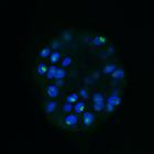
The nucleolus during mammary epithelial differentiation and early mammary tumorigenesis. Proliferating 3D cultures of MCF-10.B2 cells treated to activate ErbB2 were stained at day 20 with ANA-N antibo...
« Previous
1
...
6
7
8
9
10
11
12
13
...
263
Next »
Results per page:
10
20
50
100


")