Alternate header for print version
Advanced search
Contributors
Help
Submit
Search
menu
Cell Process
Cell Component
Cell Type
Organism
Microbial
Alzheimer's
Data Sets
Center for Research in Biological Systems
University of California, San Diego
9500 Gilman Drive
La Jolla, CA 92093-0608, USA
Voice
: (858) 534-0276
Fax
: (858) 534-7497
Email
: dorloff@ncmir.ucsd.edu
Search Results for
confocal microscopy
(2626 results)
(Not the results you were expecting?)
(Comments)
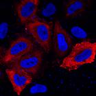
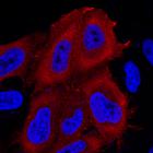
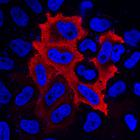
CIL:53618
NCBI Organism Classification
Homo sapiens
Biological Process
phosphorylation
Cellular Component
cell
Atypical family: The human kinome comprises 538 kinases playing essential cellular functions by catalyzing protein phosphorylations. Systematic annotation of subcellular spatial distribution of the ki...
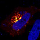
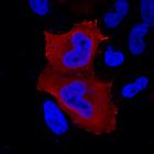
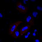
CIL:53663
NCBI Organism Classification
Homo sapiens
Biological Process
phosphorylation
Cellular Component
cell
CK1 family: The human kinome comprises 538 kinases playing essential cellular functions by catalyzing protein phosphorylations. Systematic annotation of subcellular spatial distribution of the kinome ...
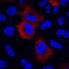
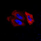
CIL:53537
NCBI Organism Classification
Homo sapiens
Biological Process
phosphorylation
Cellular Component
cell
AGC family: The human kinome comprises 538 kinases playing essential cellular functions by catalyzing protein phosphorylations. Systematic annotation of subcellular spatial distribution of the kinome ...
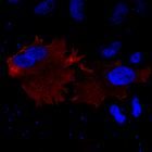
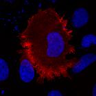
CIL:53546
NCBI Organism Classification
Homo sapiens
Biological Process
phosphorylation
Cellular Component
cell
AGC family: The human kinome comprises 538 kinases playing essential cellular functions by catalyzing protein phosphorylations. Systematic annotation of subcellular spatial distribution of the kinome ...
CIL:53547
NCBI Organism Classification
Homo sapiens
Biological Process
phosphorylation
Cellular Component
cell
AGC family: The human kinome comprises 538 kinases playing essential cellular functions by catalyzing protein phosphorylations. Systematic annotation of subcellular spatial distribution of the kinome ...
CIL:53562
NCBI Organism Classification
Homo sapiens
Biological Process
phosphorylation
Cellular Component
cell
AGC family: The human kinome comprises 538 kinases playing essential cellular functions by catalyzing protein phosphorylations. Systematic annotation of subcellular spatial distribution of the kinome ...
CIL:53564
NCBI Organism Classification
Homo sapiens
Biological Process
phosphorylation
Cellular Component
cell
AGC family: The human kinome comprises 538 kinases playing essential cellular functions by catalyzing protein phosphorylations. Systematic annotation of subcellular spatial distribution of the kinome ...
CIL:53470
NCBI Organism Classification
Homo sapiens
Biological Process
phosphorylation
Cellular Component
cell
AGC family: The human kinome comprises 538 kinases playing essential cellular functions by catalyzing protein phosphorylations. Systematic annotation of subcellular spatial distribution of the kinome ...
CIL:53484
NCBI Organism Classification
Homo sapiens
Biological Process
phosphorylation
Cellular Component
cell
AGC family: The human kinome comprises 538 kinases playing essential cellular functions by catalyzing protein phosphorylations. Systematic annotation of subcellular spatial distribution of the kinome ...
CIL:53497
NCBI Organism Classification
Homo sapiens
Biological Process
phosphorylation
Cellular Component
cell
AGC family: The human kinome comprises 538 kinases playing essential cellular functions by catalyzing protein phosphorylations. Systematic annotation of subcellular spatial distribution of the kinome ...
« Previous
1
...
137
138
139
140
141
142
143
144
...
263
Next »
Results per page:
10
20
50
100