Alternate header for print version
Advanced search
Contributors
Help
Submit
Search
menu
Cell Process
Cell Component
Cell Type
Organism
Microbial
Alzheimer's
Data Sets
Center for Research in Biological Systems
University of California, San Diego
9500 Gilman Drive
La Jolla, CA 92093-0608, USA
Voice
: (858) 534-0276
Fax
: (858) 534-7497
Email
: dorloff@ncmir.ucsd.edu
Search Results for
endoplasmic reticulum organization
(2513 results)
(Not the results you were expecting?)
(Comments)
CIL:13573
NCBI Organism Classification
Homo sapiens
Biological Process
Golgi organization
Cellular Component
Golgi stack
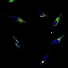
After EGF stimulation for 4 h, Helix Pomatia Lectin (HPL) (green) in HeLa cells expressing wild-type Arf1-GFP (Arf1(WT)-GFP) (gray) is redistributed from Golgi (Giantin) (red) to ER. HPL binds various...
CIL:13543
NCBI Organism Classification
Homo sapiens
Biological Process
Golgi organization
Cellular Component
Golgi stack
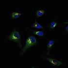
Helix Pomatia Lectin (HPL) (green) staining is redistributed out of the Golgi of HeLa cells after EGF treatment for 4 h, while the Golgi marker Giantin (red) is unaffected. HPL binds various glycans b...
CIL:13549
NCBI Organism Classification
Homo sapiens
Biological Process
Golgi organization
Cellular Component
Golgi stack
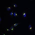
In unstimulated HeLa cells, GalNac-T1 (gray) and Helix Pomatia Lectin (HPL) (green) colocalize exclusively at the Giantin-stained Golgi apparatus (red). The Tn antigen refers to terminal α-linked N-a...
CIL:13550
NCBI Organism Classification
Homo sapiens
Biological Process
Golgi organization
Cellular Component
Golgi stack
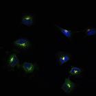
After EGF treatment of HeLa cells for 4h, a significant amount of GalNac-T1 staining (gray) is apparent in punctate and diffuse cytoplasmic cellular structures that stain positively for Helix Pomatia ...
CIL:13552
NCBI Organism Classification
Homo sapiens
Biological Process
Golgi organization
Cellular Component
Golgi stack
Helix Pomatia Lectin (HPL) (green) colocalizes at the Golgi (Giantin) (red) in unstimulated HeLa cells. The image also shows GalNac-T1 staining (gray). The Tn antigen refers to terminal α-linked N-ac...
CIL:13176
NCBI Organism Classification
Micrasterias denticulata
Biological Process
secondary cell wall biogenesis
Cellular Component
plasma membrane
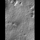
The P-face of the plasma membrane of a semicell engaged in the synthesis of the secondary wall. Hexagonal arrays of rosettes of varying size and shape can be observed. The large circular indentation...
CIL:35123
NCBI Organism Classification
Caenorhabditis elegans
Biological Process
mitosis
Cellular Component
nucleus
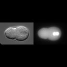
This time lapse series illustrates the early division up to the four-cell stage of a C. elegans embryo expressing YFP::CDC-48 by DIC and fluorescence microscopy. YFP::CDC-48, like YFP::UFD-1 and YFP::...
CIL:8052
NCBI Organism Classification
Homo sapiens
Biological Process
mitotic metaphase
Cellular Component
kinetochore
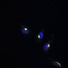
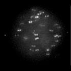
Metaphase Kinetochore Dynamics: In this HeLa cell, which is expressing both a kinetochore marker (EGFP-CENP-B) and a centrosome marker (Venus-centrin), one can visualize the dynamic nature of kinetoch...
CIL:8053
NCBI Organism Classification
Homo sapiens
Biological Process
metaphase plate congression
Cellular Component
kinetochore
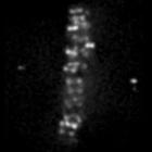
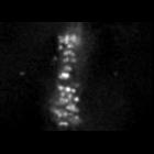
This HeLa cell is expressing two fluorescent proteins, including EGFP-CENP-B, a fluorescent protein that marks kinetochores (seen as paired dots oriented parallel to the x-axis and attached to opposit...
CIL:8057
NCBI Organism Classification
Homo sapiens
Biological Process
mitotic anaphase
Cellular Component
kinetochore
In this HeLa cell, which is expressing both a kinetochore marker (EGFP-CENP-B) and a centrosome marker (Venus-centrin), one can visualize the dynamic nature of kinetochore movements during metaphase a...
« Previous
1
...
244
245
246
247
248
249
250
251
252
Next »
Results per page:
10
20
50
100