Alternate header for print version
Advanced search
Contributors
Help
Submit
Search
menu
Cell Process
Cell Component
Cell Type
Organism
Microbial
Alzheimer's
Data Sets
Center for Research in Biological Systems
University of California, San Diego
9500 Gilman Drive
La Jolla, CA 92093-0608, USA
Voice
: (858) 534-0276
Fax
: (858) 534-7497
Email
: dorloff@ncmir.ucsd.edu
Search Results for
boundaries between regions with dif...
(1055 results)
(Not the results you were expecting?)
(Comments)
CIL:11973
NCBI Organism Classification
Thyone briareus
Biological Process
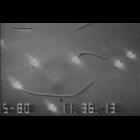
fertilization
Cellular Component
acrosome
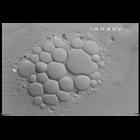
The movie shows, in real time, the explosive acrosomal reaction of Thyone (sea cucumber) sperm. The field shows several sperm heads (bright contrast) with tails of some in the plane of focus. From the...
CIL:10414
NCBI Organism Classification
Paramecium multimicronucleatum
Biological Process
cellular water homeostasis
Cellular Component
contractile vacuole
DVD quality video of the Contractile Vacuole Complex of Paramecium multimicronucleatum. In vitro CVC rounding/relaxing cycles. An in vitro CV in a ruptured Paramecium cell is surrounded by cytoplasmic...
CIL:37335
NCBI Organism Classification
none specified
Biological Process
cell migration
Cellular Component
nucleus
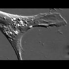
A more fully differentiated epidermal cell. The nucleus and nucleoli are on the left. The video shows the intracellular movements of lysosomes and mitochondria. The lysosomes are the small refractile ...
CIL:34848
NCBI Organism Classification
Homo sapiens
Biological Process
mitosis
Cellular Component
none specified
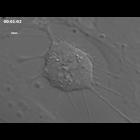
Time lapse movie of cell growth and division in cultured hTERT-RPE1 cells (telomerase immortalized human retinal pigment epithelium) using DIC optics. This movie and the stack of images used to prepar...
CIL:35188
NCBI Organism Classification
Danio rerio
Biological Process
anatomical structure morphogenesis
Cellular Component
none specified
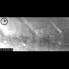
The lateral line primordium is a slug-like mass of cells that crawls down the flank of the fish, leaving clusters of cells that will form the lateral line neuromast organs. Elapsed time indicated by t...
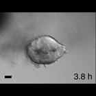
CIL:42164
NCBI Organism Classification
Homo sapiens
Biological Process
dissemination
Cellular Component
cell surface
Time-lapse movie a human tumor fragment grown in collagen I that was freed and reembedded in Matrigel. Images collected every 15 minutes and bar is 50 microns. CIL 42162 - CIL 42165 are related movi...
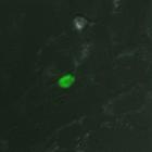
CIL:10468
NCBI Organism Classification
Toxoplasma gondii RH
Biological Process
regulation of cell shape
Cellular Component
cortical microtubule
A monolayer of human foreskin fibroblast cells was infected with Toxoplasma gondii expressing EGFP-tagged Tgbeta3-tubulin. Images overlaid with DIC. Images acquired using API DeltaVision on an Olympus...
CIL:34898
NCBI Organism Classification
none specified
Biological Process
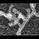
branching of actin filaments
Cellular Component
actin cytoskeleton
Improved visualization of actin filament branching in lamellipodia. EM of keratocyte or fibroblast lamellipodial actin network after cytochalasin D treatment (0.2 μM for 30 min or 0.5 μM for 10 min)...
CIL:35060
NCBI Organism Classification
none specified
Biological Process
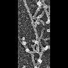
branching of actin filaments
Cellular Component
actin cytoskeleton
Electron micrograph of keratocyte or fibroblast lamellipodial actin network after unprotected extraction. All examples demonstrate frequent branching of actin filaments. Image corresponds to a singl...
CIL:39034
NCBI Organism Classification
phytoplankton
Biological Process
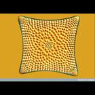
unicellular algae organization
Cellular Component
frustule
This Nomarski interference microscopy shows a marine centric diatom (unicellular alga). Diatoms are photosynthesising algae that live in most aquatic environments, both fresh and salt water. They are ...
« Previous
1
...
12
13
14
15
16
17
18
19
...
106
Next »
Results per page:
10
20
50
100


")