Alternate header for print version
Advanced search
Contributors
Help
Submit
Search
menu
Cell Process
Cell Component
Cell Type
Organism
Microbial
Alzheimer's
Data Sets
Center for Research in Biological Systems
University of California, San Diego
9500 Gilman Drive
La Jolla, CA 92093-0608, USA
Voice
: (858) 534-0276
Fax
: (858) 534-7497
Email
: dorloff@ncmir.ucsd.edu
Search Results for
phalloidin
(354 results)
(Not the results you were expecting?)
(Comments)
CIL:10216
NCBI Organism Classification
Rattus
Biological Process
developmental process
Cellular Component
cytoskeleton
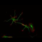
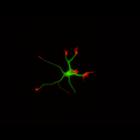
This multi-layer image shows the spatial relationship between filamentous actin (red) and microtubule array (green) in cultured hippocampal neurons, grown for 1 day in vitro. Actin staining (with rho...
CIL:26538
NCBI Organism Classification
Mus musculus
Biological Process
regulation of cell migration
Cellular Component
lamellipodium
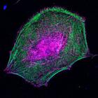
To study the molecular mechanism by which nonmuscle myosin II (MII) regulates protrusion and adhesion dynamics in migrating cells, NIH3T3 cells were transfected with myc-tagged myosin light chain (MLC...
CIL:37911
NCBI Organism Classification
Homo sapiens
Biological Process
apoptotic process
Cellular Component
cortical actin cytoskeleton
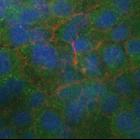
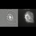
Apoptotic cells produce and transmit the bioactive lipid sphingosine-1-phosphate (S1P) during extrusion. This set of images shows that extrusion is blocked, as well as accumulation of S1P around the d...
CIL:37912
NCBI Organism Classification
Homo sapiens
Biological Process
apoptotic process
Cellular Component
cortical actin cytoskeleton
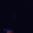
Apoptotic cells produce and transmit the bioactive lipid sphingosine-1-phosphate (S1P) during extrusion. In this confocal Z-series, an antagonist to the S1P receptor, S1P2, was used to plock signaling...
CIL:8784
NCBI Organism Classification
Rattus
Biological Process
developmental process
Cellular Component
cytoskeleton
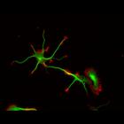
This color combined image shows the spatial relationship between filamentous actin (red) and microtubule array (green) in cultured hippocampal neurons, grown for 1 day in vitro. Actin staining (with ...
CIL:8788
NCBI Organism Classification
Rattus
Biological Process
developmental process
Cellular Component
cytoskeleton
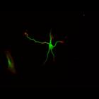
This color combined image shows the spatial relationship between filamentous actin (red) and microtubule array (green) in cultured hippocampal neurons, grown for 1 day in vitro. Actin staining (with ...
CIL:8776
NCBI Organism Classification
Rattus
Biological Process
developmental process
Cellular Component
cytoskeleton
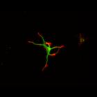
This color combined image shows the spatial relationship between filamentous actin (red) and microtubule array (green) in cultured hippocampal neurons, grown for 1 day in vitro. Actin staining (with ...
CIL:8779
NCBI Organism Classification
Rattus
Biological Process
developmental process
Cellular Component
cytoskeleton
This color combined image shows the spatial relationship between filamentous actin (red) and microtubule array (green) in cultured hippocampal neurons, grown for 1 day in vitro. Actin staining (with ...
CIL:8781
NCBI Organism Classification
Rattus
Biological Process
developmental process
Cellular Component
cytoskeleton
This color combined image shows the spatial relationship between filamentous actin (red) and microtubule array (green) in cultured hippocampal neurons, grown for 1 day in vitro. Actin staining (with ...
CIL:12017
NCBI Organism Classification
Hypsophrys nicaraguensis
Biological Process
actin axle formation
Cellular Component
actin cytoskeleton
Formation of an actin axle during the maturation of polarity. During maturation of the polarized form, the circular actin bands around the cell body transitioned to an actin axle along the cell rear. ...
« Previous
1
...
26
27
28
29
30
31
32
33
...
36
Next »
Results per page:
10
20
50
100