Alternate header for print version
Advanced search
Contributors
Help
Submit
Search
menu
Cell Process
Cell Component
Cell Type
Organism
Microbial
Alzheimer's
Data Sets
Center for Research in Biological Systems
University of California, San Diego
9500 Gilman Drive
La Jolla, CA 92093-0608, USA
Voice
: (858) 534-0276
Fax
: (858) 534-7497
Email
: dorloff@ncmir.ucsd.edu
Search Results for
primary antibody plus labeled secon...
(2148 results)
(Not the results you were expecting?)
(Comments)
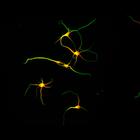
CIL:2887
NCBI Organism Classification
Rattus
Biological Process
dendrite development
Cellular Component
microtubule cytoskeleton
Development of the axon and dendritic arbors in cultured hippocampal neurons after 2 days in vitro. MAP2 staining (red) highlights the dendrites, while microtubule staining (green) reveals both the ax...
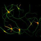
CIL:2986
NCBI Organism Classification
Rattus
Biological Process
dendrite development
Cellular Component
microtubule cytoskeleton
Development of the axon and dendritic arbors in cultured hippocampal neurons after 5 days in vitro. MAP2 staining (red) highlights the dendrites, while microtubule staining (green) reveals both the ax...
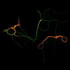
CIL:3074
NCBI Organism Classification
Rattus
Biological Process
dendrite development
Cellular Component
microtubule cytoskeleton
Development of the axon and dendritic arbors in cultured hippocampal neurons after 7 days in vitro. MAP2 staining (red) highlights the dendrites, while microtubule staining (green) reveals both the ax...
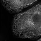
CIL:35279
NCBI Organism Classification
Rattus rattus
Biological Process
interphase
Cellular Component
microtubule organizing center
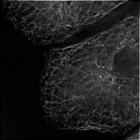
Primary rat hepatocytes in culture fixed and stained for beta-tubulin. The microtubules form a network throughout the cytoplasm. In this image they are seen originating from a brighter and denser re...
CIL:35280
NCBI Organism Classification
Rattus rattus
Biological Process
interphase
Cellular Component
microtubule organizing center
Primary rat hepatocytes in culture fixed and stained for beta-tubulin. The microtubules form a network throughout the cytoplasm. In this image they are seen originating from a brighter and denser re...
CIL:34904
NCBI Organism Classification
none specified
Biological Process
branching of actin filaments
Cellular Component
actin cytoskeleton
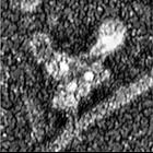
Localization of Arp2/3 complex at actin filament branching points. Xenopus keratocytes and fibroblasts were treated with CD (0.2 μM for 30 min or 0.5 μM for 10 min), extracted in the presence of pha...
CIL:34912
NCBI Organism Classification
none specified
Biological Process
branching of actin filaments
Cellular Component
actin cytoskeleton
Localization of Arp2/3 complex at actin filament branching points. Xenopus keratocytes and fibroblasts were treated with CD (0.2 μM for 30 min or 0.5 μM for 10 min), extracted in the presence of pha...
CIL:34916
NCBI Organism Classification
none specified
Biological Process
branching of actin filaments
Cellular Component
actin cytoskeleton
Localization of Arp2/3 complex at actin filament branching points. Xenopus keratocytes and fibroblasts were treated with CD (0.2 μM for 30 min or 0.5 μM for 10 min), extracted in the presence of pha...
CIL:34926
NCBI Organism Classification
Homo sapiens
Biological Process
branching of actin filaments
Cellular Component
actin cytoskeleton
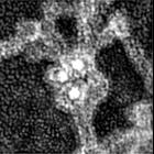
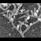
Localization of cross-linking proteins in fibroblast cytoskeleton. Immuno-EM of the cell edge or interior (CIL 34929) of cytochalasin D-treated human 356 fibroblasts stained with ABP-280 primary anti...
CIL:34927
NCBI Organism Classification
Xenopus laevis
Biological Process
branching of actin filaments
Cellular Component
actin cytoskeleton
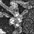
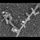
Localization of cross-linking proteins in fibroblast cytoskeleton. Immuno-EM of the cell edge or interior (CIL 34929) of cytochalasin D-treated Xenopus fibroblasts stained with α-actinin primary ant...
« Previous
1
...
10
11
12
13
14
15
16
17
...
215
Next »
Results per page:
10
20
50
100