Alternate header for print version
Advanced search
Contributors
Help
Submit
Search
menu
Cell Process
Cell Component
Cell Type
Organism
Microbial
Alzheimer's
Data Sets
Center for Research in Biological Systems
University of California, San Diego
9500 Gilman Drive
La Jolla, CA 92093-0608, USA
Voice
: (858) 534-0276
Fax
: (858) 534-7497
Email
: dorloff@ncmir.ucsd.edu
Search Results for
visualization of contiguous regions
(2856 results)
(Not the results you were expecting?)
(Comments)
CIL:34897
NCBI Organism Classification
none specified
Biological Process
branching of actin filaments
Cellular Component
actin cytoskeleton
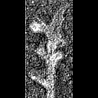
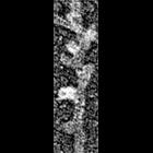
Improved visualization of actin filament branching in lamellipodia. EM of keratocyte or fibroblast lamellipodial actin network after cytochalasin D treatment (0.2 μM for 30 min or 0.5 μM for 10 min)...
CIL:34885
NCBI Organism Classification
Xenopus laevis
Biological Process
branching of actin filaments
Cellular Component
actin cytoskeleton
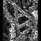
Multiple branching of actin filaments in lamellipodia of Xenopus keratocytes. This image show an enlargement of a local region from the overview of the leading edge, CIL 24786. Image corresponds to F...
CIL:34890
NCBI Organism Classification
none specified
Biological Process
branching of actin filaments
Cellular Component
actin cytoskeleton
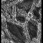
Multiple branching of actin filaments in lamellipodia of vertebrate fibroblasts. This image shows a local enlargement of the leading edge shown in overview in CIL 24788. Image corresponds to Figure 1...
CIL:35062
NCBI Organism Classification
none specified
Biological Process
branching of actin filaments
Cellular Component
actin cytoskeleton
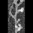
Electron micrograph of keratocyte or fibroblast lamellipodial actin network after unprotected extraction. All examples demonstrate frequent branching of actin filaments. Image corresponds to a singl...
CIL:35063
NCBI Organism Classification
none specified
Biological Process
branching of actin filaments
Cellular Component
actin cytoskeleton
Electron micrograph of keratocyte or fibroblast lamellipodial actin network after unprotected extraction. All examples demonstrate frequent branching of actin filaments. Image corresponds to a singl...
CIL:38724
NCBI Organism Classification
Chlamydomonas reinhardtii
Biological Process
photosynthesis
Cellular Component
chloroplast
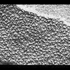
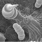
Isolated chloroplasts of the unicellular photosynthetic alga Chlamydomonas reinhardtii were cryofixed, freeze-fractured, and surface replicas observed by transmission electron microscopy. This close-u...
CIL:39116
NCBI Organism Classification
Hordeum vulgare subsp. vulgare
Biological Process
C4 photosynthesis
Cellular Component
chloroplast
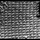
High magnification freeze-fracture image of a chloroplast from barley (Hordeum vulgare)showing the occasional repeating array of tetrameric particles. Other images in this group show the more typical ...
CIL:39117
NCBI Organism Classification
Hordeum vulgare subsp. vulgare
Biological Process
C4 photosynthesis
Cellular Component
chloroplast
High magnification freeze-fracture image of a chloroplast from barley (Hordeum vulgare)mutant lacking chlorophyll b showing an example of the occasional repeating array of tetrameric particles. Other ...
CIL:39343
NCBI Organism Classification
Helianthus annuus
Biological Process
respiratory gaseous exchange
Cellular Component
none specified
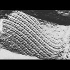
Scanning electron microscope image of sunflower lower leaf surface. Note the stomata (pore used for gas exchange) near the center of the image. This is part of an image series containing images CIL:...
CIL:40810
NCBI Organism Classification
Rattus
Biological Process
dendrite morphogenesis
Cellular Component
axon
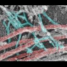
Colorized transmission electron micrograph of a platinum replica showing the cytoskeletal organization of stubby dendritic spines in extracted hippocampal neurons after 14 DIV. This imge shows branche...
« Previous
1
...
218
219
220
221
222
223
224
225
...
286
Next »
Results per page:
10
20
50
100