Alternate header for print version
Advanced search
Contributors
Help
Submit
Search
menu
Cell Process
Cell Component
Cell Type
Organism
Microbial
Alzheimer's
Data Sets
Center for Research in Biological Systems
University of California, San Diego
9500 Gilman Drive
La Jolla, CA 92093-0608, USA
Voice
: (858) 534-0276
Fax
: (858) 534-7497
Email
: dorloff@ncmir.ucsd.edu
Search Results for
Structural constituent of cytoskeleton
(125 results)
(Not the results you were expecting?)
(Comments)
Still Images
Video/Animation
Z-Stack
Time Series
CIL:38863
NCBI Organism Classification
Didinium nasutum
Biological Process
cortical cytoskeleton organization
Cellular Component
cell cortex
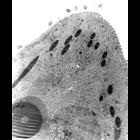
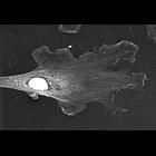
High resolution image of Didinium (non-dividing) has a massive cytopharynx at its anterior end that takes the shape of a cone-shaped proboscis when closed. This cytopharynx contains numerous lamellae ...
CIL:12302
NCBI Organism Classification
Didinium nasutum
Biological Process
cortical cytoskeleton organization
Cellular Component
cell cortex
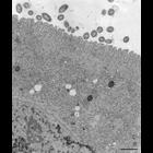
Lamellae of cytopharynx in the opisthe of a dividing cell. This cytopharynx is not yet functional and lacks most of the medium dense vesicles that accumulate between the lamellae. Toxicysts are presen...
CIL:10004
NCBI Organism Classification
Didinium nasutum
Biological Process
cortical cytoskeleton organization
Cellular Component
cell cortex
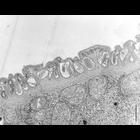
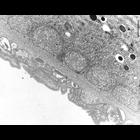
An image of the pellicle of Didinium, in cross section of a non-dividing cell, has a plasma membrane with a distinct glycocalyx on its external surface and a flattened alveolar sac system. Under the a...
CIL:12300
NCBI Organism Classification
Didinium nasutum
Biological Process
cortical cytoskeleton organization
Cellular Component
cell cortex
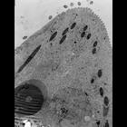
Didinium (non-dividing) has a massive cytopharynx at its anterior end that takes the shape of a cone-shaped proboscis when closed. This cytopharynx contains numerous lamellae composed of sets of micro...
CIL:10005
NCBI Organism Classification
Didinium nasutum
Biological Process
cortical cytoskeleton organization
Cellular Component
cell cortex
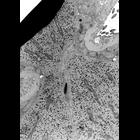
Didinium nasutum. A tangential view of the surface of a non-dividing cell shows several ribbons of microtubules between the alveolar sac and the epiplasm. The thick fibrous layer associated with the e...
CIL:38865
NCBI Organism Classification
Didinium nasutum
Biological Process
cell division
Cellular Component
cell division site
The division furrow of a dividing Didinium. A thick layer of epiplasm coated on the cytosolic side by mitochondria forms the constricting ring in the furrow. As usual mucocysts lie between the epiplas...
CIL:24784
NCBI Organism Classification
Xenopus laevis
Biological Process
actin filament organization
Cellular Component
lamellipodium
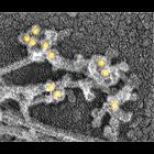
Localization of XAC (Xenopus ADF/cofilin) in Xenopus fibroblasts. Immuno-EM with XAC antibody at low magnification. High magnification view is available at CIL 24785. Nucleus and surrounding regions...
CIL:34925
NCBI Organism Classification
Xenopus laevis
Biological Process
branching of actin filaments
Cellular Component
actin cytoskeleton
Localization of cross-linking proteins in fibroblast cytoskeleton. Immuno-EM of the cell edge or interior (CIL 34928) of cytochalasin D-treated Xenopus fibroblasts stained with p21 (Arp2/3) primary a...
CIL:10454
NCBI Organism Classification
Toxoplasma gondii RH
Biological Process
regulation of cell shape
Cellular Component
cortical microtubule, transverse to long axis
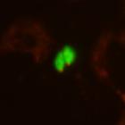
Toxoplasma gondii double stable clone expressing TgAPR1-mCherryFP (an MTOC protein) and EGFP-beta1-tubulin. 3D stack image of a live unfixed specimen acquired using API Delta Vision on Olympus ix70 w...
CIL:10542
NCBI Organism Classification
Toxoplasma gondii RH
Biological Process
regulation of cell shape
Cellular Component
cortical microtubule
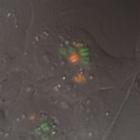
A monolayer of human foreskin fibroblast cells was infected with Toxoplasma gondii expressing YFP-tagged TgIMC1 (innermembrane complex scaffold protein) and mCherry-tagged human alpha1tubulin. Images ...
« Previous
1
...
3
4
5
6
7
8
9
10
...
13
Next »
Results per page:
10
20
50
100