Alternate header for print version
Advanced search
Contributors
Help
Submit
Search
menu
Cell Process
Cell Component
Cell Type
Organism
Microbial
Alzheimer's
Data Sets
Center for Research in Biological Systems
University of California, San Diego
9500 Gilman Drive
La Jolla, CA 92093-0608, USA
Voice
: (858) 534-0276
Fax
: (858) 534-7497
Email
: dorloff@ncmir.ucsd.edu
Search Results for
columnar/cuboidal epithelial cell
(1212 results)
(Not the results you were expecting?)
(Comments)
CIL:11199
NCBI Organism Classification
Rattus
Biological Process
cell-cell adhesion
Cellular Component
zonula adherens
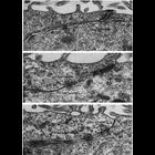
These examples of ependymal epithelium from the rat brain represent epithelia that lack zonulae occludentes. Further, there appears to be no consistent order to the junctional specializations that are...
CIL:11529
NCBI Organism Classification
Pongo pygmaeus
Biological Process
pigmentation
Cellular Component
melanosome
Figure 297 from Chapter 11 (Melanin Pigment) of 'The Cell, 2nd Ed.' by Don W. Fawcett M.D. Epidermal melanocyte from facial skin of an orangutan illustrates the interstitial location of a melanocyte ...
CIL:35996
NCBI Organism Classification
Rattus
Biological Process
secretory granule organization
Cellular Component
secretory granule
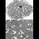
Figure 375 from Chapter 15 (Cytoplasmic Inclusions) of 'The Cell, 2nd Ed.' by Don W. Fawcett M.D. Freeze-fracture replica of a cell from rat adrenal medulla. Using this technique, secretory vesicles...
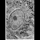
CIL:32023
NCBI Organism Classification
Xenopus laevis
Biological Process
cytokinesis
Cellular Component
nucleus
Neural plate of Xenopus laevis at early neural tube closure.
CIL:36053
NCBI Organism Classification
Mammalia
Biological Process
regulation of cell shape
Cellular Component
microtubule
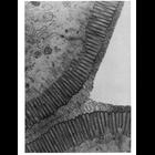
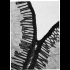
Figures 419 (upper) and 420 (lower) from Chapter 16 (Cytoplasmic matrix and cytoskeleton) of 'The Cell, 2nd Ed.' by Don W. Fawcett M.D. Electron micrographs of pillar cells of the mammalian organ of ...
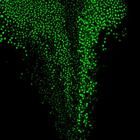
CIL:39469
NCBI Organism Classification
Xenopus laevis
Biological Process
oocyte construction
Cellular Component
nucleus
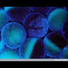
Fluorescent micrograph of stage V-VI Xenopus laevis oocytes surrounded by thousands of follicle cells, as visualized by Hoechst staining.
CIL:36002
NCBI Organism Classification
Rattus
Biological Process
secretory granule organization
Cellular Component
secretory granule
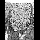
Figure 384 from Chapter 15 (Cytoplasmic Inclusions) of 'The Cell, 2nd Ed.' by Don W. Fawcett M.D. Surface mucous cells from gastric mucosa of the rat. In some species, the secretory granules from th...
CIL:36003
NCBI Organism Classification
Cavia porcellus
Biological Process
secretory granule organization
Cellular Component
secretory granule
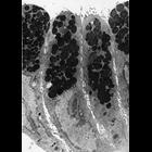
Figure 385 from Chapter 15 (Cytoplasmic Inclusions) of 'The Cell, 2nd Ed.' by Don W. Fawcett M.D. The secretory granules from surface mucous cells of the guinea pig stomach are large and pleiomorphic...
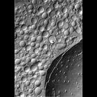
CIL:10929
NCBI Organism Classification
Felis catus
Biological Process
extracellular structure organization
Cellular Component
plasma membrane
This electron micrograph shows a region of the brush border of the cat intestine. Tufted and branched polysaccharide filaments, each a few nanometers thick, extend from the microvilli to make up the ...
CIL:10932
NCBI Organism Classification
Felis catus
Biological Process
extracellular structure organization
Cellular Component
plasma membrane
This electron micrograph highlights a darkly-stained glycocalyx rim of the brush border of the intestinal epithelium of the cat, stained en bloc with colloidal thorium. The glycocalyx is composed of ...
« Previous
1
2
3
4
5
6
7
8
9
...
122
Next »
Results per page:
10
20
50
100