Alternate header for print version
Advanced search
Contributors
Help
Submit
Search
menu
Cell Process
Cell Component
Cell Type
Organism
Microbial
Alzheimer's
Data Sets
Center for Research in Biological Systems
University of California, San Diego
9500 Gilman Drive
La Jolla, CA 92093-0608, USA
Voice
: (858) 534-0276
Fax
: (858) 534-7497
Email
: dorloff@ncmir.ucsd.edu
Search Results for
Paramecium multimicronucleatum
(292 results)
(Not the results you were expecting?)
(Comments)
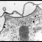
CIL:36747
NCBI Organism Classification
Paramecium multimicronucleatum
Biological Process
digestive system process
Cellular Component
food vacuole
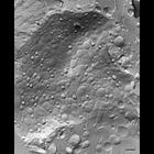
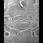
A freeze-fracture image of a DV-II transforming into a DV-III by rapid fusion of lysosomes with its membrane. The P-face shown here of the early DV-III has abundant IMPs as well as many inward blebs (...
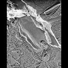
CIL:36748
NCBI Organism Classification
Paramecium multimicronucleatum
Biological Process
digestive system process
Cellular Component
food vacuole
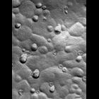
Detail of fractured early Digestion Vacuole-III membrane showing the many IMPs on the P-fracture faces of the lysosomal membranes that had just fused with the DV. The membrane will smooth out as the p...
CIL:36752
NCBI Organism Classification
Paramecium multimicronucleatum
Biological Process
digestive system process
Cellular Component
phagolysosome
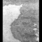
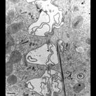
Following digestion, that only occurs in the phagolysosome (DV-III), the membrane of these DVs undergoes extensive tubulation. These tubules are ~45nm in diameter and arise from the cytosolic surface ...
CIL:36753
NCBI Organism Classification
Paramecium multimicronucleatum
Biological Process
digestive system process
Cellular Component
phagolysosome
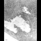
The tubules arising from late DV-III expand at their distal ends into rounded shapes. These expanded ends contain a glycocalyx like the secondary lysosomes and large IMPs also like secondary lysosomes...
CIL:36639
NCBI Organism Classification
Paramecium multimicronucleatum
Biological Process
cytoplasm organization
Cellular Component
peroxisome
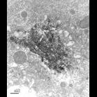
In quick-freeze deep-etch micrographs it is not easy to distinguish peroxisomes from surrounding organelles. The organelle at the top of this micrograph may be a peroxisome based on its finely granula...
CIL:36641
NCBI Organism Classification
Paramecium multimicronucleatum
Biological Process
lysosome organization
Cellular Component
crystalloid
This micrograph shows a crystal surrounded with secondary lysosomes. This was taken from a cell returned to growth medium following starvation for several days (0 day of rejuvenation). This could repr...
CIL:36648
NCBI Organism Classification
Paramecium multimicronucleatum
Biological Process
nuclear pore distribution
Cellular Component
nuclear pore
A freeze-fractured view of the nuclear envelope. The fracture in the middle of the figure exposes the cytosolic facing membrane. This can be distinguished by a fractured cytoplasmic organelle at the t...
CIL:36655
NCBI Organism Classification
Paramecium multimicronucleatum
Biological Process
conjugation
Cellular Component
cell-cell contact zone
Conjugating cells come together during or following meiosis and exchange promicronuclei. The cells actually fuse together at ridge tips and openings are formed between the two cells. TEM taken on 6/6/...
CIL:36601
NCBI Organism Classification
Paramecium multimicronucleatum
Biological Process
plasma membrane organization
Cellular Component
pellicle
This view shows a transverse ridge cut with its sloping side lying tangentially in the section plane. The granulo-fibrillar filled peaks are prominent. Striated bands appear sectioned closer to their ...
CIL:36607
NCBI Organism Classification
Paramecium multimicronucleatum
Biological Process
plasma membrane organization
Cellular Component
plasma membrane
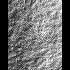
In quick-freeze deep-etch preparations the plasma membrane can be seen to contain plaques or plates of intramembrane particles (IMPs) that are regularly aligned in rows with a center-to-center spacing...
« Previous
1
...
12
13
14
15
16
17
18
19
...
30
Next »
Results per page:
10
20
50
100