Alternate header for print version
Advanced search
Contributors
Help
Submit
Search
menu
Cell Process
Cell Component
Cell Type
Organism
Microbial
Alzheimer's
Data Sets
Center for Research in Biological Systems
University of California, San Diego
9500 Gilman Drive
La Jolla, CA 92093-0608, USA
Voice
: (858) 534-0276
Fax
: (858) 534-7497
Email
: dorloff@ncmir.ucsd.edu
Search Results for
confocal microscopy
(2626 results)
(Not the results you were expecting?)
(Comments)
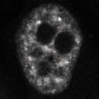
CIL:26590
NCBI Organism Classification
Homo sapiens
Biological Process
RNA splicing
Cellular Component
pre-mRNA
HeLa cells stably expressing BAC-encoded GFP-labeled snRNP protein U1-70K and imaged witb a PicoQuant MicroTime 200 confocal microscope using an Olympus 60x 1.2NA water immersion objective lens. The ...
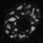
CIL:26595
NCBI Organism Classification
Homo sapiens
Biological Process
RNA splicing
Cellular Component
pre-mRNA
HeLa cells stably expressing BAC-encoded GFP-labeled snRNP protein U1-70K and treated with isoginkgetin. The protein localizes to enlarged splicing factor compartments. Cells imaged witb a Leica SP5 ...
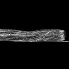
CIL:35287
NCBI Organism Classification
Lilium formosanum
Biological Process
pollen tube growth
Cellular Component
actin filament
Fixed Lilium pollen tubes were treated with fluorescent phalloidin to label filamentous actin, and confocal images obtained. A strongly labeled actin fringe near the growing tip is present. Other i...
CIL:35288
NCBI Organism Classification
Lilium formosanum
Biological Process
pollen tube growth
Cellular Component
plant-type vacuole lumen
Living Lilium pollen tube labeled with dichloro-flourescein diacetate to mark the vacuole and imaged using laser scanning focal microscopy. Shown is a central 1.0 micron thick x-y slice.The growing ...
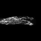
CIL:35290
NCBI Organism Classification
Lilium formosanum
Biological Process
pollen tube growth
Cellular Component
microtubule
Freeze substituted Lilium pollen tube immunolabeled for microtubules (blue>magenta) and imaged using laser scanning confocal microscopy. Shown is a projection of x-y slices revealing the distribution ...
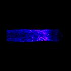
CIL:38902
NCBI Organism Classification
Mus musculus
Biological Process
neuron differentiation
Cellular Component
none specified
Confocal micrograph of neural stem cells transplanted into mouse brain Mouse neural stem cells, labelled with green fluorescent protein, have been transplanted into the brain of a newborn mouse and ar...
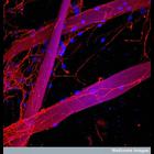
CIL:38921
NCBI Organism Classification
none specified
Biological Process
axon regeneration
Cellular Component
neuron projection
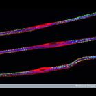
Confocal micrograph showing nerve cells growing along fibers (purple) made from a specially modified silk that is similar to that made by spiders and silkworms. Schwann cells, whose nuclei are shown i...
CIL:39103
NCBI Organism Classification
Drosophila
Biological Process
innervation
Cellular Component
neuromuscular junction
Confocal micrograph of an intact Drosophila larva imaged through the translucent cuticle showing the innervation of the dorsal (towards the back) muscle fibres by motor nerves. The muscles have been g...
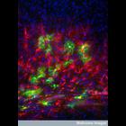
CIL:38962
NCBI Organism Classification
none specified
Biological Process
sciatic nerve fiber organization
Cellular Component
neurofilament
Confocal micrograph of teased sciatic nerve fibers. The fibers are triple labelled for neurofilament (blue), S100 (red) and dystrophin-related protein (DRP2) (green). The S100 protein is expressed in...
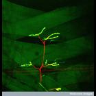
CIL:38969
NCBI Organism Classification
none specified
Biological Process
neuromuscular junction development
Cellular Component
synapse
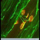
Confocal image of axons (green) making contact with muscle fibers at neuromuscular junctions. As seen here more than one nerve fiber initially contacts each muscle fiber. As development proceeds, the ...
« Previous
1
...
7
8
9
10
11
12
13
14
...
263
Next »
Results per page:
10
20
50
100