Alternate header for print version
Advanced search
Contributors
Help
Submit
Search
menu
Cell Process
Cell Component
Cell Type
Organism
Microbial
Alzheimer's
Data Sets
Center for Research in Biological Systems
University of California, San Diego
9500 Gilman Drive
La Jolla, CA 92093-0608, USA
Voice
: (858) 534-0276
Fax
: (858) 534-7497
Email
: dorloff@ncmir.ucsd.edu
Search Results for
cell-cell junction organization
(11647 results)
(Not the results you were expecting?)
(Comments)
CIL:10950
NCBI Organism Classification
Felis catus
Biological Process
plasma membrane organization
Cellular Component
basement membrane
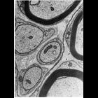
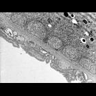
Schwann cells in peripheral nerves secrete a layer similar to the basal lamina called the lamina externa, boundary layer, or basement membrane. In this micrograph, the lamina externa surrounds the Sc...
CIL:24735
NCBI Organism Classification
Homo sapiens
Biological Process
ribosome biogenesis
Cellular Component
nucleolus
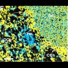
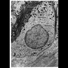
Thin section showing nucleolus of a Human neuroblastoma SK-N-SH cell imaged with a FEI Tecnai 20 energy filtered electron microscope at 200 KV and a 20 eV energy window. Overlay of net phosphorus dist...
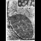
CIL:12639
NCBI Organism Classification
Paramecium multimicronucleatum
Biological Process
cytoplasm organization
Cellular Component
cytoplasm
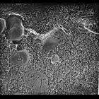
High resolution view of an early endosome with at least two coated evaginations showing, lies next to the proximal end of a fractured basal body. Other clathrin cages may indicate other evaginations f...
CIL:11050
NCBI Organism Classification
Cavia porcellus
Biological Process
nucleus organization
Cellular Component
nuclear envelope
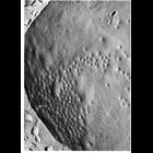
Transmission electron micrograph of surface of nuclear envelope of guinea pig spermatocyte prepared by the Freeze-fracture technique shows the unusual clustering of nuclear pores in these cells. From...
CIL:36247
NCBI Organism Classification
Vorticella convallaria
Biological Process
digestive system process
Cellular Component
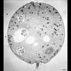
cell
11 micrographs of views of a serially-sectioned contracted Vorticella convallaria cell that show the main features of this cell. This figure shows 5 food vacuoles and the edges of 2 more. The connecti...
CIL:11021
NCBI Organism Classification
Homo sapiens
Biological Process
nucleus organization
Cellular Component
nuclear chromosome
Transmission electron micrograph of isolated HeLa cell metaphase chromosome decondensed by removal of histones with dextran sulfate and heparin, centrifuged onto a carbon film and stained with uranyl ...
CIL:11056
NCBI Organism Classification
Hirudinea
Biological Process
nucleus organization
Cellular Component
nuclear envelope
Transmission electron micrograph illustrating the unusually prominent fibrous nuclear lamina (arrows) seen in many invertebrates such as this leech ganglion cell. From Fawcett (1966) Am J. Anat. 119:...
CIL:38862
NCBI Organism Classification
Didinium nasutum
Biological Process
cortical cytoskeleton organization
Cellular Component
cell cortex
A high resolution view of a dividing Didinium nasutum. This large ciliate is capable of engulfing Paramecium. It has a thick ectoplasm containing numerous mucocysts embedded in its extensive epiplasm....
CIL:9928
NCBI Organism Classification
Didinium nasutum
Biological Process
cortical cytoskeleton organization
Cellular Component
cell cortex
A high resolution tangential view of the surface of a non-dividing cell shows several ribbons of microtubules between the alveolar sac and the epiplasm. The thick fibrous layer associated with the epi...
CIL:11528
NCBI Organism Classification
Homo sapiens
Biological Process
pigmentation
Cellular Component
melanosome
Figure 296 from Chapter 11 (Melanin Pigment) of 'The Cell, 2nd Ed.' by Don W. Fawcett M.D. Melanocyte at the dermoepidermal junction, flanked by portions of two adjacent epithelial cells from African...
« Previous
1
...
12
13
14
15
16
17
18
19
...
1165
Next »
Results per page:
10
20
50
100