Alternate header for print version
Advanced search
Contributors
Help
Submit
Search
menu
Cell Process
Cell Component
Cell Type
Organism
Microbial
Alzheimer's
Data Sets
Center for Research in Biological Systems
University of California, San Diego
9500 Gilman Drive
La Jolla, CA 92093-0608, USA
Voice
: (858) 534-0276
Fax
: (858) 534-7497
Email
: dorloff@ncmir.ucsd.edu
Search Results for
detection of electrons
(1490 results)
(Not the results you were expecting?)
(Comments)
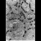
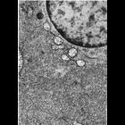
CIL:11137
NCBI Organism Classification
Mammalia
Biological Process
pinocytosis
Cellular Component
vesicle
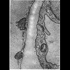
Capillary endothelial cells from mammalian cardiac muscle caught in the act of fluid-phase micropinocytosis. Arrows indicate multiple sites where invaginaton along the membrane suggests vesicles formi...
CIL:11214
NCBI Organism Classification
Aphrodita aculeata
Biological Process
cell adhesion
Cellular Component
desmosome
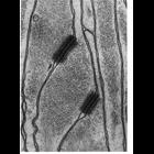
Glial cells in the nerve cord of the annelid worm, Aphrodita aculeata are rich in 10 nm filaments, and desmosomes are abundant. Figure 89 from Chapter 3 (Junctional Specializations) of 'The Cell, 2nd ...
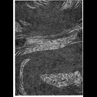
CIL:11235
NCBI Organism Classification
none specified
Biological Process
cell communication
Cellular Component
gap junction
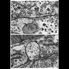
Upper: a gap junction on the boundary between two hepatic cells shows a typical narrow and straight interface. Lower: in some preparations, gap junctions appear curved, and project into one of the ce...
CIL:11358
NCBI Organism Classification
Ovis aries
Biological Process
post-translational protein modification
Cellular Component
Golgi apparatus
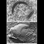
Figures 197 (upper) and 198 (lower) from Chapter 6 (Golgi Apparatus) of 'The Cell, 2nd Ed.' by Don W. Fawcett M.D. show electron microscopic views of the Golgi complex. Upper panel: A thin section pr...
CIL:11362
NCBI Organism Classification
Mus musculus
Biological Process
post-translational protein modification
Cellular Component
Golgi apparatus
Figure 199 from Chapter 6 (Golgi Apparatus) of 'The Cell, 2nd Ed.' by Don W. Fawcett M.D. The Golgi apparatus in epithelial cells from the mouse epididymis. The convex, or cis, side of the Golgi sho...
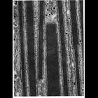
CIL:10765
NCBI Organism Classification
Myotis lucifugus
Biological Process
translation
Cellular Component
endoplasmic reticulum
Figure 169 from Chapter 5 (Endoplasmic Reticulum) of 'The Cell, 2nd Ed.' by Don W. Fawcett M.D. Electron micrograph of rough endoplasmic reticulum (RER) in an acinar cell from the pancreas of the sma...
CIL:10779
NCBI Organism Classification
Batrachoseps attenuatus
Biological Process
translation
Cellular Component
endoplasmic reticulum
Figure 178 from Chapter 5 (Endoplasmic Reticulum) of 'The Cell, 2nd Ed.' by Don W. Fawcett M.D. Electron micrograph of a liver cell from the slender salamander, Batrachoseps attenuatus. Here, high l...
CIL:10796
NCBI Organism Classification
Phodopus
Biological Process
glycogen biosynthetic process
Cellular Component
endoplasmic reticulum
Figure 183 from Chapter 5 (Endoplasmic Reticulum) of 'The Cell, 2nd Ed.' by Don W. Fawcett M.D. Electron micrograph of an hepatic liver cell from the hamster. The area of the cell shown is dense wit...
CIL:10800
NCBI Organism Classification
Didelphimorphia
Biological Process
cholesterol biosynthetic process
Cellular Component
endoplasmic reticulum
Figure 187 from Chapter 5 (Endoplasmic Reticulum) of 'The Cell, 2nd Ed.' by Don W. Fawcett M.D. An abundance of smooth endoplasmic reticulum is apparent in this Leydig cell of the opossum testes, ass...
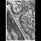
CIL:10946
NCBI Organism Classification
Siphonaptera
Biological Process
extracellular structure organization
Cellular Component
basal lamina
This electron micrograph shows the basal lamina along the midgut epithelium of the flea. Unlike the vertebrate basal lamina, which appears as a homogenous layer, the basal lamina in insects often app...
« Previous
1
...
14
15
16
17
18
19
20
21
...
149
Next »
Results per page:
10
20
50
100