Alternate header for print version
Advanced search
Contributors
Help
Submit
Search
menu
Cell Process
Cell Component
Cell Type
Organism
Microbial
Alzheimer's
Data Sets
Center for Research in Biological Systems
University of California, San Diego
9500 Gilman Drive
La Jolla, CA 92093-0608, USA
Voice
: (858) 534-0276
Fax
: (858) 534-7497
Email
: dorloff@ncmir.ucsd.edu
Search Results for
Embryonic cell
(11655 results)
(Not the results you were expecting?)
(Comments)
CIL:39791
NCBI Organism Classification
Lytechinus pictus
Biological Process
embryonic morphogenesis
Cellular Component
extracellular matrix
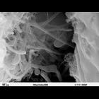
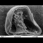
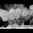
Scanning electron microscope image of Strongylocentrotus drobachiensus [sea urchin] embryo at the late gastrula stage. This high magnification image of the embryo shows the primary mesenchyme syncytia...
CIL:39787
NCBI Organism Classification
Lytechinus pictus
Biological Process
embryonic morphogenesis
Cellular Component
filopodium
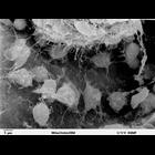
Scanning electron microscope image of Strongylocentrotus drobachiensus [sea urchin] at the gastrula stage. Embryo was split open to reveal the blastocoel cavity. There are several migrating mesenchyme...
CIL:37411
NCBI Organism Classification
Danio rerio
Biological Process
anatomical structure morphogenesis
Cellular Component
cytoplasm
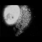
Zebrafish embryo at 80% epiboly. The initial frame shows the junction of the enveloping layer with the yolk cell. The next frame is at a focal plane deeper within the embryo. The blastoderm margin is ...
CIL:39790
NCBI Organism Classification
Lytechinus pictus
Biological Process
embryonic morphogenesis
Cellular Component
cell surface
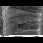
Scanning electron microscope image of Strongylocentrotus drobachiensus [sea urchin] gastrula. This is a higher magnification image of CIL39765. The embryo was split open to reveal a nice cross-section...
CIL:39784
NCBI Organism Classification
Lytechinus pictus
Biological Process
cell migration
Cellular Component
cell surface
Scanning electron microscope image of Strongylocentrotus drobachiensus [sea urchin] at the gastrula stage. Embryo was cut in half to reveal blastocoel cavity, containing blastocoel matrix material, pr...
CIL:37402
NCBI Organism Classification
Danio rerio
Biological Process
anatomical structure morphogenesis
Cellular Component
yolk platelets
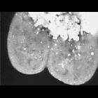
Four cell stage zebrafish embryo, stained with 100 micromolar Bodipy 505/515. In the upper portion of the image, the yolk cell contains large spherical yolk platelets. The blastomeres undergoing merob...
CCDB:3629
Species
chicken
Organ
ciliary ganglion
Cell type
ciliary ganglion neuron
System
peripheral nervous system
Structure
calycal synapse
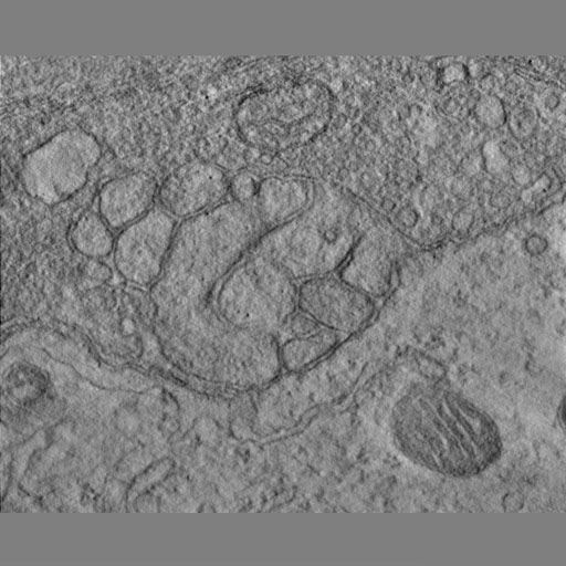
Serial tomography of embryonic ciliary ganglion spine mat
CIL:25539
NCBI Organism Classification
Drosophila melanogaster
Biological Process
heart development
Cellular Component
nucleus
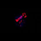
A 3D reconstruction of the cardiac outflow tract (OFT) region (horizontal and vertical rotation) from stage-16 wild-type embryo stained for Ladybird early (Lbe; blue) and Tinman (Tin; red). Heart-anch...
CIL:39789
NCBI Organism Classification
Lytechinus pictus
Biological Process
syncytial ring formation
Cellular Component
cell surface
Scanning electron microscope image of Strongylocentrotus drobachiensus [sea urchin] embryo at the late gastrula stage. This is a high magnification image of CIL 39785. Embryo was cut to reveal blastoc...
CIL:35212
NCBI Organism Classification
Mus musculus
Biological Process
cell motility
Cellular Component
ruffle
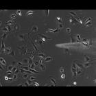
Time lapse movie of mouse embryonic fibroblasts in culture imaged at 30 second intervals by phase contrast microscopy. A micropipette is positioned near the cells to deliver either a growth factor or...
« Previous
1
...
22
23
24
25
26
27
28
29
...
1166
Next »
Results per page:
10
20
50
100


")
